3W51 | pdb_00003w51

Tankyrase in complex with 2-hydroxy-4-methylquinoline


Experimental Data Snapshot

  • Method: X-RAY DIFFRACTION
  • Resolution: 2.00 Å
  • R-Value Free: 
    0.243 (Depositor), 0.243 (DCC) 
  • R-Value Work: 
    0.187 (Depositor), 0.191 (DCC) 
  • R-Value Observed: 
    0.190 (Depositor) 

Starting Model: experimental
View more details

wwPDB Validation 3D Report Full Report

Validation slider image for 3W51

Ligand Structure Quality Assessment 


This is version 1.1 of the entry. See complete history

Literature

Fragment-based ligand design of novel potent inhibitors of tankyrases.

Larsson, E.A.Jansson, A.E.Ng, F.M.Then, S.W.Panicker, R.Liu, B.Sangthongpitag, K.Pendharkar, V.Tai, S.J.Hill, J.Dan, C.Ho, S.Y.Cheong, W.W.Poulsen, A.Blanchard, S.Lin, G.R.Alam, J.Keller, T.H.Nordlund, P.

(2013) J Med Chem 56: 4497-4508

  • DOI: https://doi.org/10.1021/jm400211f
  • Primary Citation Related Structures: 
    3W51, 4IUE, 4J1Z, 4J21, 4J22, 4J3L, 4J3M

  • PubMed Abstract: 

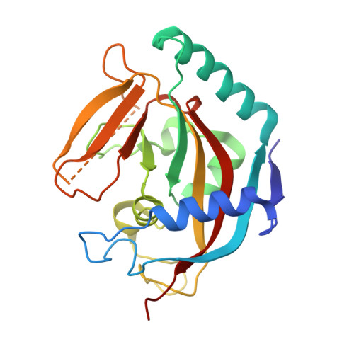
    Tankyrases constitute potential drug targets for cancer and myelin-degrading diseases. We have applied a structure- and biophysics-driven fragment-based ligand design strategy to discover a novel family of potent inhibitors for human tankyrases. Biophysical screening based on a thermal shift assay identified highly efficient fragments binding in the nicotinamide-binding site, a local hot spot for fragment binding. Evolution of the fragment hit 4-methyl-1,2-dihydroquinolin-2-one (2) along its 7-vector yields dramatic affinity improvements in the first cycle of expansion. A crystal structure of 7-(2-fluorophenyl)-4-methylquinolin-2(1H)-one (11) reveals that the nonplanar compound extends with its fluorine atom into a pocket, which coincides with a region of the active site where structural differences are seen between tankyrases and other poly(ADP-ribose) polymerase (PARP) family members. A further cycle of optimization yielded compounds with affinities and IC50 values in the low nanomolar range and with good solubility, PARP selectivity, and ligand efficiency.


  • Organizational Affiliation
    • School of Biological Sciences, Nanyang Technological University, Lab 07-01, 61 Biopolis Drive (Proteos), Singapore 138673. andreas.larsson@ntu.edu.sg

Macromolecule Content 

  • Total Structure Weight: 48.82 kDa 
  • Atom Count: 3,731 
  • Modeled Residue Count: 418 
  • Deposited Residue Count: 420 
  • Unique protein chains: 1

Macromolecules

Find similar proteins by:|  3D Structure
Entity ID: 1
MoleculeChains  Sequence LengthOrganismDetailsImage
Tankyrase-2
A, B
210Homo sapiensMutation(s): 0 
Gene Names: PARP5BTANK2TNKLTNKS2
EC: 2.4.2.30 (PDB Primary Data), 2.4.2 (UniProt)
UniProt & NIH Common Fund Data Resources
Find proteins for Q9H2K2 (Homo sapiens)
Explore Q9H2K2 
Go to UniProtKB:  Q9H2K2
PHAROS:  Q9H2K2
GTEx:  ENSG00000107854 
Entity Groups
Sequence Clusters30% Identity50% Identity70% Identity90% Identity95% Identity100% Identity
UniProt GroupQ9H2K2
Sequence Annotations
Expand
Reference Sequence

Small Molecules

Ligands 3 Unique
IDChains Name / Formula / InChI Key2D Diagram3D Interactions
AJ2

Query on AJ2


Download:Ideal Coordinates CCD File
C [auth A],
G [auth B]
4-methylquinolin-2-ol
C10 H9 N O
APLVPBUBDFWWAD-UHFFFAOYSA-N
SO4

Query on SO4


Download:Ideal Coordinates CCD File
E [auth A],
F [auth A],
I [auth B],
J [auth B]
SULFATE ION
O4 S
QAOWNCQODCNURD-UHFFFAOYSA-L
ZN

Query on ZN


Download:Ideal Coordinates CCD File
D [auth A],
H [auth B]
ZINC ION
Zn
PTFCDOFLOPIGGS-UHFFFAOYSA-N

Experimental Data & Validation

Experimental Data

  • Method: X-RAY DIFFRACTION
  • Resolution: 2.00 Å
  • R-Value Free:  0.243 (Depositor), 0.243 (DCC) 
  • R-Value Work:  0.187 (Depositor), 0.191 (DCC) 
  • R-Value Observed: 0.190 (Depositor) 
Space Group: C 2 2 21
Unit Cell:
Length ( Å )Angle ( ˚ )
a = 94.276α = 90
b = 95.038β = 90
c = 116.497γ = 90
Software Package:
Software NamePurpose
REFMACrefinement
PDB_EXTRACTdata extraction
HKL-2000data collection
MOSFLMdata reduction
SCALAdata scaling
PHASESphasing

Structure Validation

View Full Validation Report


Ligand Structure Quality Assessment 


Entry History 

Deposition Data

Revision History  (Full details and data files)

  • Version 1.0: 2013-07-10
    Type: Initial release
  • Version 1.1: 2023-11-08
    Changes: Data collection, Database references, Derived calculations, Refinement description