398D

3'-DNA-RNA-5' JUNCTION FORMED DURING INITIATION OF MINUS-STRAND SYNTHESIS OF HIV REPLICATION


Experimental Data Snapshot

  • Method: X-RAY DIFFRACTION
  • Resolution: 1.94 Å
  • R-Value Free: 0.291 
  • R-Value Work: 0.220 
  • R-Value Observed: 0.220 

wwPDB Validation   3D Report Full Report


This is version 1.4 of the entry. See complete history


Literature

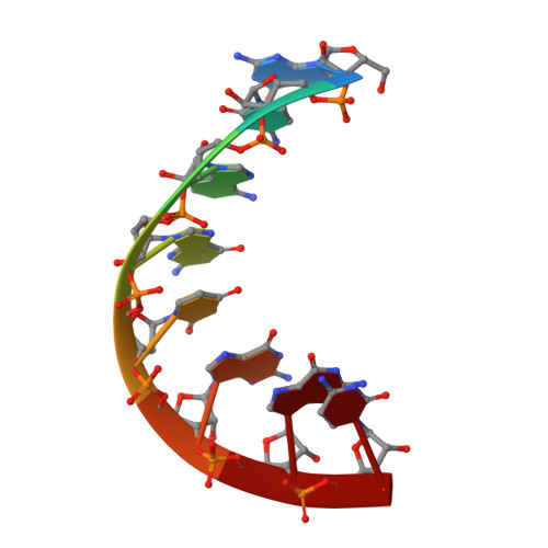
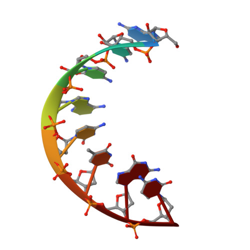
Crystal structure of an eight-base pair duplex containing the 3'-DNA-RNA-5' junction formed during initiation of minus-strand synthesis of HIV replication.

Mueller, U.Maier, G.Mochi Onori, A.Cellai, L.Heumann, H.Heinemann, U.

(1998) Biochemistry 37: 12005-12011

  • DOI: https://doi.org/10.1021/bi981152y
  • Primary Citation of Related Structures:  
    398D

  • PubMed Abstract: 

    During initiation of minus-strand synthesis by HIV-1 reverse transcriptase, a 3'-DNA-RNA-5' junction is formed involving the 3'-end of tRNAlys,3. The HIV-RT-associated RNase H cleaves the RNA template strand specifically, opposite the newly synthesized DNA strand. We have determined the crystal structure at 1.9 A resolution of an eight-base pair hybrid duplex representing the junction to identify global or local structural perturbations which may be recognized by HIV-RT RNase H. The junction octamer is in a global A-type conformation throughout. A base pair step with distinct stacking geometry and variable backbone conformation is located next to the main endonucleolytic cleavage site. This base pair step may serve as a recognition site for HIV-RT RNase H.


  • Organizational Affiliation

    Forschungsgruppe Kristallographie, Max-Delbrück-Centrum für Molekulare Medizin, Berlin, Germany.


Macromolecules

Find similar nucleic acids by:  Sequence   |   3D Structure  

Entity ID: 1
MoleculeChains LengthOrganismImage
RNA (5'-R(*GP*CP*AP*GP*UP*GP*GP*C)-3')
A, C
8N/A
Sequence Annotations
Expand
  • Reference Sequence

Find similar nucleic acids by:  Sequence   |   3D Structure  

Entity ID: 2
MoleculeChains LengthOrganismImage
DNA/RNA (5'-R(*GP*CP*CP*AP)-D(*CP*TP*GP*C)-3')
B, D
8N/A
Sequence Annotations
Expand
  • Reference Sequence
Experimental Data & Validation

Experimental Data

  • Method: X-RAY DIFFRACTION
  • Resolution: 1.94 Å
  • R-Value Free: 0.291 
  • R-Value Work: 0.220 
  • R-Value Observed: 0.220 
  • Space Group: P 21 21 21
Unit Cell:
Length ( Å )Angle ( ˚ )
a = 42.74α = 90
b = 44.15β = 90
c = 51.93γ = 90
Software Package:
Software NamePurpose
AMoREphasing
X-PLORrefinement
DENZOdata reduction
SCALEPACKdata scaling

Structure Validation

View Full Validation Report


Entry History 

Deposition Data

Revision History  (Full details and data files)

  • Version 1.0: 1998-10-06
    Type: Initial release
  • Version 1.1: 2008-05-22
    Changes: Version format compliance
  • Version 1.2: 2011-07-13
    Changes: Version format compliance
  • Version 1.3: 2024-02-21
    Changes: Data collection, Database references
  • Version 1.4: 2024-04-03
    Changes: Refinement description