391D | pdb_0000391d

STRUCTURAL VARIABILITY AND NEW INTERMOLECULAR INTERACTIONS OF Z-DNA IN CRYSTALS OF D(PCPGPCPGPCPG)


Experimental Data Snapshot

  • Method: X-RAY DIFFRACTION
  • Resolution: 2.75 Å
  • R-Value Free: 
    0.238 (Depositor) 
  • R-Value Work: 
    0.199 (Depositor), 0.199 (DCC) 
  • R-Value Observed: 
    0.199 (Depositor) 

wwPDB Validation 3D Report Full Report

Validation slider image for 391D

This is version 1.4 of the entry. See complete history

Literature

Structural variability and new intermolecular interactions of Z-DNA in crystals of d(pCpGpCpGpCpG).

Malinina, L.Tereshko, V.Ivanova, E.Subirana, J.A.Zarytova, V.Nekrasov, Y.

(1998) Biophys J 74: 2482-2490

  • DOI: https://doi.org/10.1016/S0006-3495(98)77956-1
  • Primary Citation Related Structures: 
    390D, 391D, 392D

  • PubMed Abstract: 

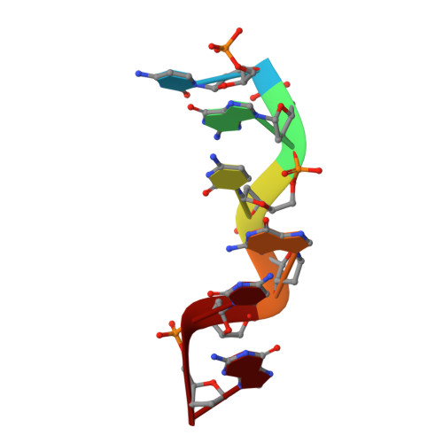
    We have determined the single crystal x-ray structure of the synthetic DNA hexamer d(pCpGpCpGpCpG) in two different crystal forms. The hexamer pCGCGCG has the Z-DNA conformation and in both cases the asymmetric unit contains more than one Z-DNA duplex. Crystals belong to the space group C222(1) with a = 69.73, b = 52.63, and c = 26.21 A, and to the space group P2(1) with a = 49.87, b = 41.26, c = 21.91 A, and gamma = 97.12 degrees. Both crystals show new crystal packing modes. The molecules also show striking new features when compared with previously determined Z-DNA structures: 1) the bases in one duplex have a large inclination with respect to the helical axis, which alters the overall shape of the molecule. 2) Some cytosine nitrogens interact by hydrogen bonding with phosphates in neighbor molecules. Similar base-phosphate interactions had been previously detected in some B-DNA crystals. 3) Basepair stacking between the ends of neighbor molecules is variable and no helical continuity is maintained between contiguous hexamer duplexes.


  • Organizational Affiliation
    • Engelhardt Institute of Molecular Biology RAS, Moscow, Russia. lucy@crystal.eimb.rssi.ru

Macromolecule Content 

  • Total Structure Weight: 10.86 kDa 
  • Atom Count: 761 
  • Modeled Residue Count: 36 
  • Deposited Residue Count: 36 
  • Unique nucleic acid chains: 1

Macromolecules

Find similar nucleic acids by:  Sequence
Entity ID: 1
MoleculeChains LengthOrganismImage
DNA (5'-D(P*CP*GP*CP*GP*CP*G)-3')
A, B, C, D, E
A, B, C, D, E, F
6N/A
Sequence Annotations
Expand
Reference Sequence

Experimental Data & Validation

Experimental Data

  • Method: X-RAY DIFFRACTION
  • Resolution: 2.75 Å
  • R-Value Free:  0.238 (Depositor) 
  • R-Value Work:  0.199 (Depositor), 0.199 (DCC) 
  • R-Value Observed: 0.199 (Depositor) 
Space Group: P 1 1 21
Unit Cell:
Length ( Å )Angle ( ˚ )
a = 49.87α = 90
b = 41.26β = 90
c = 21.91γ = 97.12
Software Package:
Software NamePurpose
X-PLORrefinement
SYNTEXdata reduction
SYNTEXdata scaling

Structure Validation

View Full Validation Report


Entry History 

Deposition Data

Revision History  (Full details and data files)

  • Version 1.0: 1998-05-05
    Type: Initial release
  • Version 1.1: 2008-05-22
    Changes: Version format compliance
  • Version 1.2: 2011-07-13
    Changes: Version format compliance
  • Version 1.3: 2018-04-04
    Changes: Data collection
  • Version 1.4: 2024-02-21
    Changes: Data collection, Database references