A novel end-to-end binding of two netropsins to the DNA decamers d(CCCCCIIIII)2, d(CCCBr5CCIIIII)2and d(CBr5CCCCIIIII)2.
Chen, X., Mitra, S.N., Rao, S.T., Sekar, K., Sundaralingam, M.(1998) Nucleic Acids Res 26: 5464-5471
- PubMed: 9826773 Search on PubMedSearch on PubMed Central
- DOI: https://doi.org/10.1093/nar/26.23.5464
- Primary Citation Related Structures:
358D, 375D, 474D - PubMed Abstract:
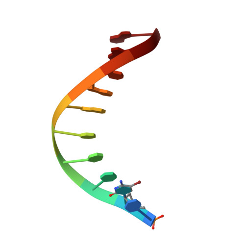
Netropsin is bound to the DNA decamer d(CCCCCIIIII)2, the C-4 bromo derivative d(CCCBr5CCIIIII)2and the C-2 bromo derivative d(CBr5CCCCIIIII)2in a novel 2:1 mode. Complexes of the native decamer and the C-4 bromo derivative are isomorphous, space group P1, unit cell dimensions a = 32.56 A (32.66), b = 32.59 A (32.77), c = 37.64 A (37.71), alpha = 86.30 degrees (86.01 degrees), beta = 84.50 degrees (84.37 degrees), gamma = 68.58 degrees (68.90 degrees) with two independent molecules (A and B) in the asymmetric unit (values in parentheses are for the derivative). The C-2 bromo derivative is hexagonal P61, unit cell dimensions a = b = 32.13 A, c = 143.92, gamma = 120 degrees with one molecule in the asymmetric unit. The structures were solved by the molecular replacement method. The novelty of the structures is that there are two netropsins bound end-to-end in the minor groove of each B-DNA decamer which has nearly a complete turn. The netropsins are held by hydrogen bonding interactions to the base atoms and by sandwiching van der Waal's interactions from the sugar-phosphate backbones of the double helix similar to every other drug.DNA complex. Each netropsin molecule spans approximately 5 bp. The netropsins refined with their guanidinium heads facing each other at the center, although an orientational disorder for the netropsins cannot be excluded. The amidinium ends stretch out toward the junctions and bind to the adjacent duplexes in the columns of stacked symmetry-related complexes. Both cationic ends of netropsin are bridged by water molecules in one of the independent molecules (molecule A) of the triclinic structures and also the hexagonal structure to form pseudo-continuous drug.decamer helices.
- The Ohio State University, Biological Macromolecular Structure Center, Departments of Chemistry and Biochemistry, 012 Rightmire Hall, 1060 Carmack Road, Columbus, OH 43210, USA.
Organizational Affiliation: