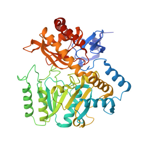
2ZUK

The crystal structure of alpha-amino-epsilon-caprolactam racemase from Achromobacter obae complexed with epsilon caprolactam (different binding mode)


Experimental Data Snapshot

  • Method: X-RAY DIFFRACTION
  • Resolution: 2.41 Å
  • R-Value Free: 0.251 
  • R-Value Work: 0.191 
  • R-Value Observed: 0.194 

Starting Model: experimental
View more details

wwPDB Validation   3D Report Full Report


Ligand Structure Quality Assessment 


This is version 1.2 of the entry. See complete history


Literature

The novel structure of a pyridoxal 5'-phosphate-dependent fold-type I racemase, alpha-amino-epsilon-caprolactam racemase from Achromobacter obae

Okazaki, S.Suzuki, A.Mizushima, T.Kawano, T.Komeda, H.Asano, Y.Yamane, T.

(2009) Biochemistry 48: 941-950

  • DOI: https://doi.org/10.1021/bi801574p
  • Primary Citation of Related Structures:  
    2ZUK, 3DXV, 3DXW

  • PubMed Abstract: 

    Alpha-amino-epsilon-caprolactam (ACL) racemase (ACLR) from Achromobacter obae catalyzes the interconversion of l- and d-ACL. ACLR belongs to the fold-type I group of pyridoxal 5'-phosphate (PLP) dependent enzymes. In this study, the first crystal structures of a fold-type I racemase are solved for the native form and epsilon-caprolactam-complexed form of ACLR at 2.21 and 2.40 A resolution, respectively. Based on the location of epsilon-caprolactam in the complex structure, the substrate-binding site is assigned between Trp49 and Tyr137. The carboxyl group of Asp210 is a reasonable candidate that recognizes the nitrogen atom of a lactam or amide in the substrate. Based on a structural comparison with fold-type III alanine racemase, Tyr137 is potentially the acid/base catalytic residue that is essential for the two-base racemization mechanism. The overall structure of ACLR is similar to that of fold-type I enzymes. A structural comparison with these enzymes explains the different reaction specificities.


  • Organizational Affiliation

    Department of Biotechnology, Graduate School of Engineering, Nagoya University, Chikusa, Nagoya 464-8603, Japan.


Macromolecules
Find similar proteins by:  (by identity cutoff)  |  3D Structure
Entity ID: 1
MoleculeChains Sequence LengthOrganismDetailsImage
Alpha-amino-epsilon-caprolactam racemase
A, B
439Achromobacter obaeMutation(s): 0 
EC: 5.1.1.15
UniProt
Find proteins for Q7M181 (Achromobacter obae)
Explore Q7M181 
Go to UniProtKB:  Q7M181
Entity Groups  
Sequence Clusters30% Identity50% Identity70% Identity90% Identity95% Identity100% Identity
UniProt GroupQ7M181
Sequence Annotations
Expand
  • Reference Sequence
Experimental Data & Validation

Experimental Data

  • Method: X-RAY DIFFRACTION
  • Resolution: 2.41 Å
  • R-Value Free: 0.251 
  • R-Value Work: 0.191 
  • R-Value Observed: 0.194 
  • Space Group: P 1 21 1
Unit Cell:
Length ( Å )Angle ( ˚ )
a = 56.581α = 90
b = 60.653β = 103.07
c = 105.315γ = 90
Software Package:
Software NamePurpose
REFMACrefinement
HKL-2000data collection
HKL-2000data reduction
HKL-2000data scaling
MOLREPphasing

Structure Validation

View Full Validation Report


Ligand Structure Quality Assessment 


Entry History 

Deposition Data

Revision History  (Full details and data files)

  • Version 1.0: 2009-02-17
    Type: Initial release
  • Version 1.1: 2011-07-13
    Changes: Non-polymer description, Version format compliance
  • Version 1.2: 2023-11-01
    Changes: Data collection, Database references, Derived calculations, Refinement description