2ZUA | pdb_00002zua

Crystal structure of nucleoside diphosphate kinase from Haloarcula quadrata


Experimental Data Snapshot

  • Method: X-RAY DIFFRACTION
  • Resolution: 2.59 Å
  • R-Value Free: 
    0.289 (Depositor), 0.275 (DCC) 
  • R-Value Work: 
    0.222 (Depositor), 0.213 (DCC) 
  • R-Value Observed: 
    0.225 (Depositor) 

Starting Model: experimental
View more details

wwPDB Validation 3D Report Full Report

Validation slider image for 2ZUA

This is version 1.2 of the entry. See complete history

Literature

Molecular mechanism of distinct salt-dependent enzyme activity of two halophilic nucleoside diphosphate kinases

Yamamura, A.Ichimura, T.Kamekura, M.Mizuki, T.Usami, R.Makino, T.Ohtsuka, J.Miyazono, K.Okai, M.Nagata, K.Tanokura, M.

(2009) Biophys J 96: 4692-4700

  • DOI: https://doi.org/10.1016/j.bpj.2009.03.012
  • Primary Citation Related Structures: 
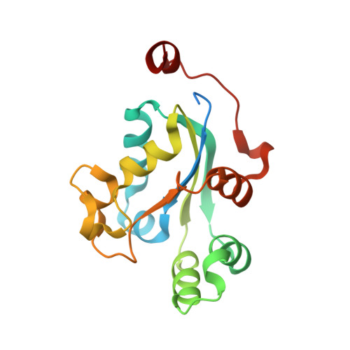
    2ZUA

  • PubMed Abstract: 

    Nucleoside diphosphate kinases from haloarchaea Haloarcula quadrata (NDK-q) and H. sinaiiensis (NDK-s) are identical except for one out of 154 residues, i.e., Arg(31) in NDK-q and Cys(31) in NDK-s. However, the salt-dependent activity profiles of NDK-q and NDK-s are quite different: the optimal NaCl concentrations of NDK-q and NDK-s are 1 M and 2 M, respectively. We analyzed the relationships of the secondary, tertiary, and quaternary structures and NDK activity of these NDKs at various salt concentrations, and revealed that 1), NDK-q is present as a hexamer under a wide range of salt concentrations (0.2-4 M NaCl), whereas NDK-s is present as a hexamer at an NaCl concentration above 2 M and as a dimer at NaCl concentrations below 1 M; 2), dimeric NDK-s has lower activity than hexameric NDK-s; and 3), dimeric NDK-s has higher helicity than hexameric NDK-s. We also determined the crystal structure of hexameric NDK-q, and revealed that Arg(31) plays an important role in stabilizing the hexamer. Thus the substitution of Arg (as in NDK-q) to Cys (as in NDK-s) at position 31 destabilizes the hexameric assembly, and causes dissociation to less active dimers at low salt concentrations.


  • Organizational Affiliation
    • Department of Applied Biological Chemistry, Graduate School of Agricultural and Life Sciences, University of Tokyo, Bunkyo-ku, Tokyo 113-8657, Japan.

Macromolecule Content 

  • Total Structure Weight: 78.95 kDa 
  • Atom Count: 4,901 
  • Modeled Residue Count: 611 
  • Deposited Residue Count: 696 
  • Unique protein chains: 1

Macromolecules

Find similar proteins by:|  3D Structure
Entity ID: 1
MoleculeChains  Sequence LengthOrganismDetailsImage
Nucleoside diphosphate kinase
A, B, C, D
174Haloarcula quadrataMutation(s): 0 
Gene Names: ndk
EC: 2.7.4.6
UniProt
Find proteins for Q401C5 (Haloarcula quadrata)
Explore Q401C5 
Go to UniProtKB:  Q401C5
Entity Groups
Sequence Clusters30% Identity50% Identity70% Identity90% Identity95% Identity100% Identity
UniProt GroupQ401C5
Sequence Annotations
Expand
Reference Sequence

Experimental Data & Validation

Experimental Data

  • Method: X-RAY DIFFRACTION
  • Resolution: 2.59 Å
  • R-Value Free:  0.289 (Depositor), 0.275 (DCC) 
  • R-Value Work:  0.222 (Depositor), 0.213 (DCC) 
  • R-Value Observed: 0.225 (Depositor) 
Space Group: P 63
Unit Cell:
Length ( Å )Angle ( ˚ )
a = 73.571α = 90
b = 73.571β = 90
c = 214.271γ = 120
Software Package:
Software NamePurpose
REFMACrefinement
HKL-2000data collection
HKL-2000data reduction
HKL-2000data scaling
MOLREPphasing

Structure Validation

View Full Validation Report


Entry History 

Deposition Data

Revision History  (Full details and data files)

  • Version 1.0: 2009-08-25
    Type: Initial release
  • Version 1.1: 2011-07-13
    Changes: Version format compliance
  • Version 1.2: 2023-11-01
    Changes: Data collection, Database references, Refinement description