Fast binding kinetics and conserved 3D structure underlie the antagonistic activity of mutant TNF: useful information for designing artificial proteo-antagonists
Mukai, Y., Nakamura, T., Yoshioka, Y., Shibata, H., Abe, Y., Nomura, T., Taniai, M., Ohta, T., Nakagawa, S., Tsunoda, S., Kamada, H., Yamagata, Y., Tsutsumi, Y.(2009) J Biochem 146: 167-172
- PubMed: 19386778
- DOI: https://doi.org/10.1093/jb/mvp065
- Primary Citation of Related Structures:
2ZPX - PubMed Abstract:
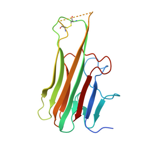
Tumour necrosis factor (TNF) is an important cytokine that induces an inflammatory response predominantly through the TNF receptor-1 (TNFR1). A crucial strategy for the treatment of many autoimmune diseases, therefore, is to block the binding of TNF to TNFR1. We previously identified a TNFR1-selective antagonistic mutant TNF (R1antTNF) from a phage library containing six randomized amino acid residues at the receptor-binding site (amino acids 84-89). Two R1antTNFs, R1antTNF-T2 (A84S, V85T, S86T, Y87H, Q88N and T89Q) and R1antTNF-T8 (A84T, V85P, S86A, Y87I, Q88N and T89R), were successfully isolated from this library. Here, we analysed R1antTNF-T8 using surface plasmon resonance spectroscopy and X-ray crystallography to determine the mechanism underlying the antagonistic activity of R1antTNF. The kinetic association/dissociation parameters of R1antTNF-T8 were higher than those of wild-type TNF, indicating more rapid bond dissociation. X-ray crystallographic analysis suggested that the binding mode of the T89R mutation changed from a hydrophobic to an electrostatic interaction, which may be responsible for the antagonistic behaviour of R1antTNF. Knowledge of these structure-function relationships will facilitate the design of novel TNF inhibitors based on the cytokine structure.
- Osaka University, Yamadaoka, Suita, Japan.
Organizational Affiliation:


