Histidine kinase regulation by a cyclophilin-like inhibitor
Jacques, D.A., Langley, D.B., Jeffries, C.M., Cunningham, K.A., Burkholder, W.F., Guss, J.M., Trewhella, J.(2008) J Mol Biol 384: 422-435
- PubMed: 18823995 Search on PubMed
- DOI: https://doi.org/10.1016/j.jmb.2008.09.017
- Primary Citation Related Structures:
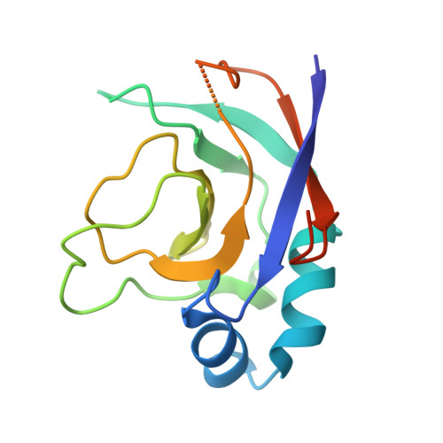
2ZP2 - PubMed Abstract:
The sensor histidine kinase A (KinA) from Bacillus subtilis triggers a phosphorelay that activates sporulation. The antikinase KipI prevents sporulation by binding KinA and inhibiting the autophosphorylation reaction. Using neutron contrast variation, mutagenesis, and fluorescence data, we show that two KipI monomers bind via their C-domains at a conserved proline in the KinA dimerization and histidine-phosphotransfer (DHp) domain. Our crystal structure of the KipI C-domain reveals the binding motif has a distinctive hydrophobic groove formed by a five-stranded antiparallel beta-sheet; a characteristic of the cyclophilin family of proteins that bind prolines and often act as cis-trans peptidyl-prolyl isomerases. We propose that the DHp domain of KinA transmits conformational signals to regulate kinase activity via this proline-mediated interaction. Given that both KinA and KipI homologues are widespread in the bacterial kingdom, this mechanism has broad significance in bacterial signal transduction.
- School of Molecular and Microbial Biosciences, University of Sydney, Sydney, New South Wales 2006, Australia.
Organizational Affiliation: