2ZOV | pdb_00002zov

Structure of the periplasmic domain of MotB from Salmonella (crystal form I)


Experimental Data Snapshot

  • Method: X-RAY DIFFRACTION
  • Resolution: 2.00 Å
  • R-Value Free: 
    0.265 (Depositor), 0.265 (DCC) 
  • R-Value Work: 
    0.233 (Depositor), 0.233 (DCC) 
  • R-Value Observed: 
    0.233 (Depositor) 

wwPDB Validation 3D Report Full Report

Validation slider image for 2ZOV

This is version 1.2 of the entry. See complete history

Literature

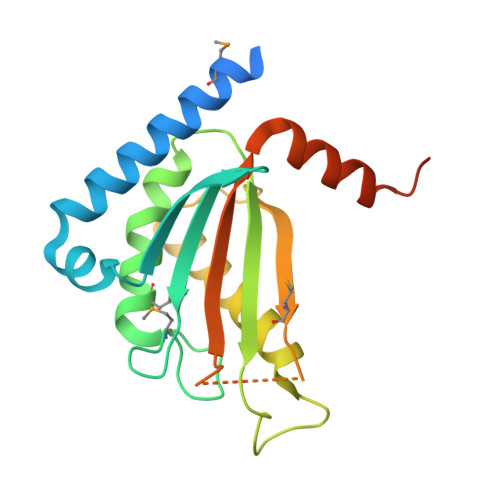
Stator assembly and activation mechanism of the flagellar motor by the periplasmic region of MotB

Kojima, S.Imada, K.Sakuma, M.Sudo, Y.Kojima, C.Minamino, T.Homma, M.Namba, K.

(2009) Mol Microbiol 73: 710-718

  • DOI: https://doi.org/10.1111/j.1365-2958.2009.06802.x
  • Primary Citation Related Structures: 
    2ZOV, 2ZVY, 2ZVZ

  • PubMed Abstract: 

    Torque generation in the Salmonella flagellar motor is coupled to translocation of H(+) ions through the proton-conducting channel of the Mot protein stator complex. The Mot complex is believed to be anchored to the peptidoglycan (PG) layer by the putative peptidoglycan-binding (PGB) domain of MotB. Proton translocation is activated only when the stator is installed into the motor. We report the crystal structure of a C-terminal periplasmic fragment of MotB (MotB(C)) that contains the PGB domain and includes the entire periplasmic region essential for motility. Structural and functional analyses indicate that the PGB domains must dimerize in order to form the proton-conducting channel. Drastic conformational changes in the N-terminal portion of MotB(C) are required both for PG binding and the proton channel activation.


  • Organizational Affiliation
    • Division of Biological Science, Graduate School of Science, Nagoya University, Chikusa-Ku, Nagoya 464-8602, Japan.

Macromolecule Content 

  • Total Structure Weight: 23.98 kDa 
  • Atom Count: 1,508 
  • Modeled Residue Count: 166 
  • Deposited Residue Count: 210 
  • Unique protein chains: 1

Macromolecules

Find similar proteins by:|  3D Structure
Entity ID: 1
MoleculeChains  Sequence LengthOrganismDetailsImage
Chemotaxis protein motB210Salmonella enterica subsp. enterica serovar TyphimuriumMutation(s): 0 
UniProt
Find proteins for P55892 (Salmonella typhimurium (strain LT2 / SGSC1412 / ATCC 700720))
Explore P55892 
Go to UniProtKB:  P55892
Entity Groups
Sequence Clusters30% Identity50% Identity70% Identity90% Identity95% Identity100% Identity
UniProt GroupP55892
Sequence Annotations
Expand
Reference Sequence

Small Molecules

Modified Residues  1 Unique
IDChains TypeFormula2D DiagramParent
MSE
Query on MSE
A
L-PEPTIDE LINKINGC5 H11 N O2 SeMET

Experimental Data & Validation

Experimental Data

  • Method: X-RAY DIFFRACTION
  • Resolution: 2.00 Å
  • R-Value Free:  0.265 (Depositor), 0.265 (DCC) 
  • R-Value Work:  0.233 (Depositor), 0.233 (DCC) 
  • R-Value Observed: 0.233 (Depositor) 
Space Group: P 32 2 1
Unit Cell:
Length ( Å )Angle ( ˚ )
a = 99.74α = 90
b = 99.74β = 90
c = 43.96γ = 120
Software Package:
Software NamePurpose
CNSrefinement
ADSCdata collection
MOSFLMdata reduction
SCALAdata scaling
SOLVEphasing

Structure Validation

View Full Validation Report


Entry History 

Deposition Data

Revision History  (Full details and data files)

  • Version 1.0: 2009-06-09
    Type: Initial release
  • Version 1.1: 2011-07-13
    Changes: Version format compliance
  • Version 1.2: 2024-11-06
    Changes: Data collection, Database references, Derived calculations, Structure summary