2ZOA

Malonate-bound structure of the glycerophosphodiesterase from Enterobacter aerogenes (GpdQ) COLLECTED AT 1.280 ANGSTROM


Experimental Data Snapshot

  • Method: X-RAY DIFFRACTION
  • Resolution: 2.40 Å
  • R-Value Free: 0.179 
  • R-Value Work: 0.159 
  • R-Value Observed: 0.160 

wwPDB Validation   3D Report Full Report


This is version 1.2 of the entry. See complete history


Literature

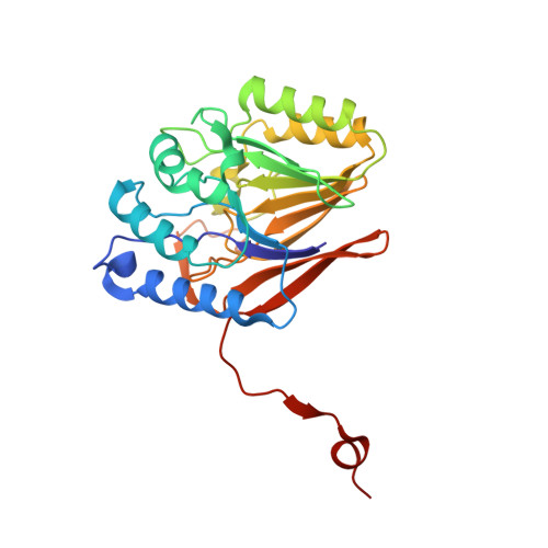
Malonate-bound structure of the glycerophosphodiesterase from Enterobacter aerogenes (GpdQ) and characterization of the native Fe2+ metal-ion preference.

Jackson, C.J.Hadler, K.S.Carr, P.D.Oakley, A.J.Yip, S.Schenk, G.Ollis, D.L.

(2008) Acta Crystallogr Sect F Struct Biol Cryst Commun 64: 681-685

  • DOI: https://doi.org/10.1107/S1744309108017600
  • Primary Citation of Related Structures:  
    2ZO9, 2ZOA

  • PubMed Abstract: 

    The structure of a malonate-bound form of the glycerophosphodiesterase from Enterobacter aerogenes, GpdQ, has been refined at a resolution of 2.2 A to a final R factor of 17.1%. The structure was originally solved to 2.9 A resolution using SAD phases from Zn2+ metal ions introduced into the active site of the apoenzyme [Jackson et al. (2007), J. Mol. Biol. 367, 1047-1062]. However, the 2.9 A resolution was insufficient to discern significant details of the architecture of the binuclear metal centre that constitutes the active site. Furthermore, kinetic analysis revealed that the enzyme lost a significant amount of activity in the presence of Zn2+, suggesting that it is unlikely to be a catalytically relevant metal ion. In this communication, a higher resolution structure of GpdQ is presented in which malonate is visibly coordinated in the active site and analysis of the native metal-ion preference is presented using atomic absorption spectroscopy and anomalous scattering. Catalytic implications of the structure and its Fe2+ metal-ion preference are discussed.


  • Organizational Affiliation

    Research School of Chemistry, Australian National University, Australia.


Macromolecules
Find similar proteins by:  (by identity cutoff)  |  3D Structure
Entity ID: 1
MoleculeChains Sequence LengthOrganismDetailsImage
Phosphohydrolase
A, B
274Klebsiella aerogenesMutation(s): 0 
Gene Names: GpdQ
EC: 3.1.4.46
UniProt
Find proteins for Q6XBH1 (Klebsiella aerogenes)
Explore Q6XBH1 
Go to UniProtKB:  Q6XBH1
Entity Groups  
Sequence Clusters30% Identity50% Identity70% Identity90% Identity95% Identity100% Identity
UniProt GroupQ6XBH1
Sequence Annotations
Expand
  • Reference Sequence
Experimental Data & Validation

Experimental Data

  • Method: X-RAY DIFFRACTION
  • Resolution: 2.40 Å
  • R-Value Free: 0.179 
  • R-Value Work: 0.159 
  • R-Value Observed: 0.160 
  • Space Group: P 21 3
Unit Cell:
Length ( Å )Angle ( ˚ )
a = 164.343α = 90
b = 164.343β = 90
c = 164.343γ = 90
Software Package:
Software NamePurpose
REFMACrefinement
HKL-2000data collection
DENZOdata reduction
SCALEPACKdata scaling
MOLREPphasing

Structure Validation

View Full Validation Report


Entry History 

Deposition Data

Revision History  (Full details and data files)

  • Version 1.0: 2008-10-07
    Type: Initial release
  • Version 1.1: 2011-07-13
    Changes: Advisory, Version format compliance
  • Version 1.2: 2023-11-01
    Changes: Data collection, Database references, Derived calculations, Refinement description