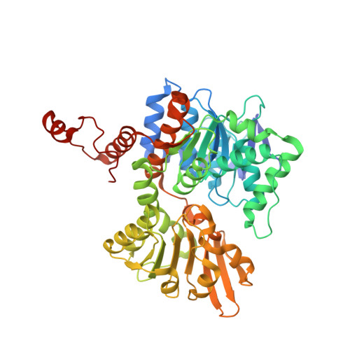
2ZJ1

Crystal structure of Mycobacterium tuberculosis S-adenosyl-L-homocysteine hydrolase in ternary complex with NAD and 3'-keto-aristeromycin


Experimental Data Snapshot

  • Method: X-RAY DIFFRACTION
  • Resolution: 2.01 Å
  • R-Value Free: 0.247 
  • R-Value Work: 0.190 
  • R-Value Observed: 0.192 

Starting Model: experimental
View more details

wwPDB Validation   3D Report Full Report


Ligand Structure Quality Assessment 


This is version 1.2 of the entry. See complete history


Literature

Crystal structures of Mycobacterium tuberculosis S-adenosyl-L-homocysteine hydrolase in ternary complex with substrate and inhibitors.

Reddy, M.C.Kuppan, G.Shetty, N.D.Owen, J.L.Ioerger, T.R.Sacchettini, J.C.

(2008) Protein Sci 17: 2134-2144

  • DOI: https://doi.org/10.1110/ps.038125.108
  • Primary Citation of Related Structures:  
    2ZIZ, 2ZJ0, 2ZJ1, 3CE6, 3DHY

  • PubMed Abstract: 

    S-adenosylhomocysteine hydrolase (SAHH) is a ubiquitous enzyme that plays a central role in methylation-based processes by maintaining the intracellular balance between S-adenosylhomocysteine (SAH) and S-adenosylmethionine. We report the first prokaryotic crystal structure of SAHH, from Mycobacterium tuberculosis (Mtb), in complex with adenosine (ADO) and nicotinamide adenine dinucleotide. Structures of complexes with three inhibitors are also reported: 3'-keto aristeromycin (ARI), 2-fluoroadenosine, and 3-deazaadenosine. The ARI complex is the first reported structure of SAHH complexed with this inhibitor, and confirms the oxidation of the 3' hydroxyl to a planar keto group, consistent with its prediction as a mechanism-based inhibitor. We demonstrate the in vivo enzyme inhibition activity of the three inhibitors and also show that 2-fluoradenosine has bactericidal activity. While most of the residues lining the ADO-binding pocket are identical between Mtb and human SAHH, less is known about the binding mode of the homocysteine (HCY) appendage of the full substrate. We report the 2.0 A resolution structure of the complex of SAHH cocrystallized with SAH. The most striking change in the structure is that binding of HCY forces a rotation of His363 around the backbone to flip out of contact with the 5' hydroxyl of the ADO and opens access to a nearby channel that leads to the surface. This complex suggests that His363 acts as a switch that opens up to permit binding of substrate, then closes down after release of the cleaved HCY. Differences in the entrance to this access channel between human and Mtb SAHH are identified.


  • Organizational Affiliation

    Department of Biochemistry and Biophysics, Texas A&M University, College Station, Texas 77843-2128, USA.


Macromolecules
Find similar proteins by:  (by identity cutoff)  |  3D Structure
Entity ID: 1
MoleculeChains Sequence LengthOrganismDetailsImage
Adenosylhomocysteinase
A, B, C, D
495Mycobacterium tuberculosis H37RvMutation(s): 0 
Gene Names: ahcYsahHRv3248cMT3346MTCY20B11.23c
EC: 3.3.1.1
UniProt
Find proteins for P9WGV3 (Mycobacterium tuberculosis (strain ATCC 25618 / H37Rv))
Explore P9WGV3 
Go to UniProtKB:  P9WGV3
Entity Groups  
Sequence Clusters30% Identity50% Identity70% Identity90% Identity95% Identity100% Identity
UniProt GroupP9WGV3
Sequence Annotations
Expand
  • Reference Sequence
Small Molecules
Ligands 2 Unique
IDChains Name / Formula / InChI Key2D Diagram3D Interactions
NAD
Query on NAD

Download Ideal Coordinates CCD File 
F [auth A],
H [auth B],
I [auth C],
L [auth D]
NICOTINAMIDE-ADENINE-DINUCLEOTIDE
C21 H27 N7 O14 P2
BAWFJGJZGIEFAR-NNYOXOHSSA-N
ARJ
Query on ARJ

Download Ideal Coordinates CCD File 
E [auth A],
G [auth B],
J [auth C],
K [auth D]
(2S,3R,5R)-3-(6-amino-9H-purin-9-yl)-2-hydroxy-5-(hydroxymethyl)cyclopentanone
C11 H13 N5 O3
CWNCBQJCRSRXGI-KCRUCZTKSA-N
Experimental Data & Validation

Experimental Data

  • Method: X-RAY DIFFRACTION
  • Resolution: 2.01 Å
  • R-Value Free: 0.247 
  • R-Value Work: 0.190 
  • R-Value Observed: 0.192 
  • Space Group: P 1 21 1
Unit Cell:
Length ( Å )Angle ( ˚ )
a = 94.854α = 90
b = 111.85β = 96.49
c = 100.403γ = 90
Software Package:
Software NamePurpose
REFMACrefinement
ADSCdata collection
HKL-2000data reduction
HKL-2000data scaling
AMoREphasing

Structure Validation

View Full Validation Report


Ligand Structure Quality Assessment 


Entry History 

Deposition Data

Revision History  (Full details and data files)

  • Version 1.0: 2008-09-16
    Type: Initial release
  • Version 1.1: 2011-07-13
    Changes: Source and taxonomy, Version format compliance
  • Version 1.2: 2023-08-30
    Changes: Data collection, Database references, Derived calculations, Refinement description