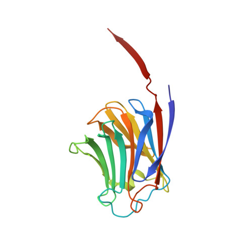
Structural basis for the tumor cell apoptosis-inducing activity of an antitumor lectin from the edible mushroom Agrocybe aegerita
Yang, N., Li, D.F., Feng, L., Xiang, Y., Liu, W., Sun, H., Wang, D.C.(2009) J Mol Biology 387: 694-705
- PubMed: 19361423
- DOI: https://doi.org/10.1016/j.jmb.2009.02.002
- Primary Citation of Related Structures:
2ZGK, 2ZGL, 2ZGM, 2ZGN, 2ZGO, 2ZGP, 2ZGQ, 2ZGR, 2ZGS, 2ZGT, 2ZGU - PubMed Abstract:
Lectin AAL (Agrocybe aegerita lectin) from the edible mushroom A. aegerita is an antitumor protein that exerts its tumor-suppressing function via apoptosis-inducing activity in cancer cells. The crystal structures of ligand-free AAL and its complex with lactose have been determined. The AAL structure shows a dimeric organization, and each protomer adopts a prototype galectin fold. To identify the structural determinants for antitumor effects arising from the apoptosis-inducing activity of AAL, 11 mutants were prepared and subjected to comprehensive investigations covering oligomerization detection, carbohydrate binding test, apoptosis-inducing activity assay, and X-ray crystallographic analysis. The results show that dimerization of AAL is a prerequisite for its tumor cell apoptosis-inducing activity, and both galactose and glucose are basic moieties of functional carbohydrate ligands for lectin bioactivity. Furthermore, we have identified a hydrophobic pocket that is essential for the protein's apoptosis-inducing activity but independent of its carbohydrate binding and dimer formation. This hydrophobic pocket comprises a hydrophobic cluster including residues Leu33, Leu35, Phe93, and Ile144, and is involved in AAL's function mechanism as an integrated structural motif. Single mutants such as F93G or I144G do not disrupt carbohydrate binding and homodimerization capabilities, but abolish the bioactivity of the protein. These findings reveal the structural basis for the antitumor property of AAL, which may lead to de novo designs of antitumor drugs based on AAL as a prototype model.
- National Laboratory of Biomacromolecules, Institute of Biophysics, Chinese Academy of Sciences, Chaoyang District, Beijing, PR China.
Organizational Affiliation:


