2ZDU | pdb_00002zdu

Crystal Structure of human JNK3 complexed with an isoquinolone inhibitor


Experimental Data Snapshot

  • Method: X-RAY DIFFRACTION
  • Resolution: 2.50 Å
  • R-Value Free: 
    0.319 (Depositor), 0.232 (DCC) 
  • R-Value Work: 
    0.241 (Depositor), 0.241 (DCC) 

Starting Model: experimental
View more details

wwPDB Validation 3D Report Full Report

Validation slider image for 2ZDU

Ligand Structure Quality Assessment 


This is version 1.2 of the entry. See complete history

Literature

Discovery, synthesis and biological evaluation of isoquinolones as novel and highly selective JNK inhibitors (1)

Asano, Y.Kitamura, S.Ohra, T.Aso, K.Igata, H.Tamura, T.Kawamoto, T.Tanaka, T.Sogabe, S.Matsumoto, S.Yamaguchi, M.Kimura, H.Itoh, F.

(2008) Bioorg Med Chem 16: 4715-4732

  • DOI: https://doi.org/10.1016/j.bmc.2008.02.027
  • Primary Citation Related Structures: 
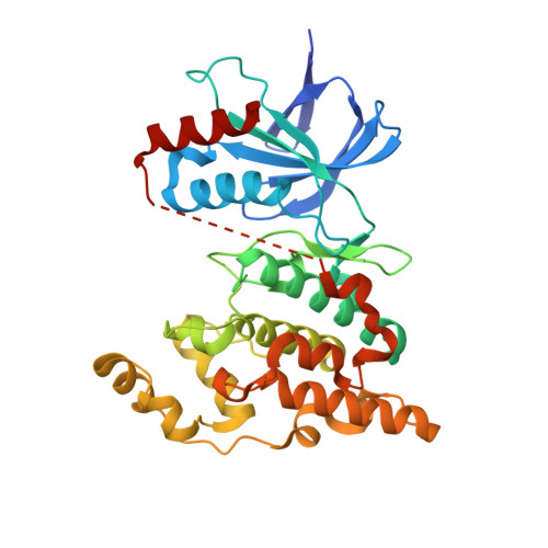
    2ZDU

  • PubMed Abstract: 

    A novel series of 4-phenylisoquinolones were synthesized and evaluated as c-Jun N-terminal kinase (JNK) inhibitors. Initial modification at the 2- and 3-positions of the isoquinolone ring of hit compound 4, identified from high-throughput screening, led to the lead compound 6b. The optimization was carried out using a JNK1-binding model of 6b and several compounds exhibited potent JNK inhibition. Among them, 11g significantly inhibited cardiac hypertrophy in rat pressure-overload models without affecting blood pressure and the concept of JNK inhibitors as novel therapeutic agents for heart failure was confirmed.


  • Organizational Affiliation
    • Medicinal Chemistry Research Laboratories, Pharmaceutical Research Division, Takeda Pharmaceutical Company, Ltd, 17-85, Jusohonmachi 2-chome, Osaka 532-8686, Japan. Asano_Yasutomi@takeda.co.jp

Macromolecule Content 

  • Total Structure Weight: 42.62 kDa 
  • Atom Count: 2,774 
  • Modeled Residue Count: 330 
  • Deposited Residue Count: 364 
  • Unique protein chains: 1

Macromolecules

Find similar proteins by:|  3D Structure
Entity ID: 1
MoleculeChains  Sequence LengthOrganismDetailsImage
Mitogen-activated protein kinase 10364Homo sapiensMutation(s): 0 
EC: 2.7.11.24
UniProt & NIH Common Fund Data Resources
Find proteins for P53779 (Homo sapiens)
Explore P53779 
Go to UniProtKB:  P53779
PHAROS:  P53779
GTEx:  ENSG00000109339 
Entity Groups
Sequence Clusters30% Identity50% Identity70% Identity90% Identity95% Identity100% Identity
UniProt GroupP53779
Sequence Annotations
Expand
Reference Sequence

Small Molecules

Ligands 1 Unique
IDChains Name / Formula / InChI Key2D Diagram3D Interactions
446

Query on 446


Download:Ideal Coordinates CCD File
B [auth A]4-[(4-{[6-bromo-3-(methoxycarbonyl)-1-oxo-4-phenylisoquinolin-2(1H)-yl]methyl}phenyl)amino]-4-oxobutanoic acid
C28 H23 Br N2 O6
UJOQZQKRUNERFC-UHFFFAOYSA-N
Binding Affinity Annotations 
IDSourceBinding Affinity
446 BindingDB:  2ZDU IC50: 9.2 (nM) from 1 assay(s)

Experimental Data & Validation

Experimental Data

  • Method: X-RAY DIFFRACTION
  • Resolution: 2.50 Å
  • R-Value Free:  0.319 (Depositor), 0.232 (DCC) 
  • R-Value Work:  0.241 (Depositor), 0.241 (DCC) 
Space Group: P 21 21 21
Unit Cell:
Length ( Å )Angle ( ˚ )
a = 56.956α = 90
b = 70.267β = 90
c = 107.947γ = 90
Software Package:
Software NamePurpose
CNXrefinement
CrystalCleardata collection
d*TREKdata reduction
d*TREKdata scaling
AMoREphasing

Structure Validation

View Full Validation Report


Ligand Structure Quality Assessment 


Entry History 

Deposition Data

Revision History  (Full details and data files)

  • Version 1.0: 2008-09-23
    Type: Initial release
  • Version 1.1: 2011-07-13
    Changes: Version format compliance
  • Version 1.2: 2023-11-01
    Changes: Data collection, Database references, Derived calculations, Refinement description