2Z86

Crystal structure of chondroitin polymerase from Escherichia coli strain K4 (K4CP) complexed with UDP-GlcUA and UDP


Experimental Data Snapshot

  • Method: X-RAY DIFFRACTION
  • Resolution: 2.40 Å
  • R-Value Free: 0.286 
  • R-Value Work: 0.225 
  • R-Value Observed: 0.225 

wwPDB Validation   3D Report Full Report


Ligand Structure Quality Assessment 


This is version 1.3 of the entry. See complete history


Literature

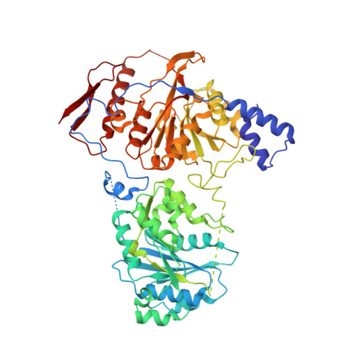
Crystal structure of chondroitin polymerase from Escherichia coli K4.

Osawa, T.Sugiura, N.Shimada, H.Hirooka, R.Tsuji, A.Shirakawa, T.Fukuyama, K.Kimura, M.Kimata, K.Kakuta, Y.

(2009) Biochem Biophys Res Commun 378: 10-14

  • DOI: https://doi.org/10.1016/j.bbrc.2008.08.121
  • Primary Citation of Related Structures:  
    2Z86, 2Z87

  • PubMed Abstract: 

    Elongation of glycosaminoglycan chains, such as heparan and chondroitin, is catalyzed by bi-functional glycosyltransferases, for which both 3-dimensional structures and reaction mechanisms remain unknown. The bacterial chondroitin polymerase K4CP catalyzes elongation of the chondroitin chain by alternatively transferring the GlcUA and GalNAc moiety from UDP-GlcUA and UDP-GalNAc to the non-reducing ends of the chondroitin chain. Here, we have determined the crystal structure of K4CP in the presence of UDP and UDP-GalNAc as well as with UDP and UDP-GlcUA. The structures consisted of two GT-A fold domains in which the two active sites were 60A apart. UDP-GalNAc and UDP-GlcUA were found at the active sites of the N-terminal and C-terminal domains, respectively. The present K4CP structures have provided the structural basis for further investigating the molecular mechanism of biosynthesis of chondroitin chain.


  • Organizational Affiliation

    Laboratory of Structural Biology, Graduate School of Systems Life Sciences, Kyushu University, Fukuoka 812-8581, Japan.


Macromolecules
Find similar proteins by:  (by identity cutoff)  |  3D Structure
Entity ID: 1
MoleculeChains Sequence LengthOrganismDetailsImage
Chondroitin synthase
A, B, C, D
625Escherichia coliMutation(s): 0 
Gene Names: kfoC
EC: 2.4.1.175 (PDB Primary Data), 2.4.1.226 (PDB Primary Data)
UniProt
Find proteins for Q8L0V4 (Escherichia coli)
Explore Q8L0V4 
Go to UniProtKB:  Q8L0V4
Entity Groups  
Sequence Clusters30% Identity50% Identity70% Identity90% Identity95% Identity100% Identity
UniProt GroupQ8L0V4
Sequence Annotations
Expand
  • Reference Sequence
Small Molecules
Ligands 3 Unique
IDChains Name / Formula / InChI Key2D Diagram3D Interactions
UGA
Query on UGA

Download Ideal Coordinates CCD File 
E [auth A]
F [auth A]
J [auth B]
N [auth C]
Q [auth D]
E [auth A],
F [auth A],
J [auth B],
N [auth C],
Q [auth D],
R [auth D]
URIDINE-5'-DIPHOSPHATE-GLUCURONIC ACID
C15 H22 N2 O18 P2
HDYANYHVCAPMJV-LXQIFKJMSA-N
UDP
Query on UDP

Download Ideal Coordinates CCD File 
I [auth B],
M [auth C]
URIDINE-5'-DIPHOSPHATE
C9 H14 N2 O12 P2
XCCTYIAWTASOJW-XVFCMESISA-N
MN
Query on MN

Download Ideal Coordinates CCD File 
G [auth A]
H [auth A]
K [auth B]
L [auth B]
O [auth C]
G [auth A],
H [auth A],
K [auth B],
L [auth B],
O [auth C],
P [auth C],
S [auth D],
T [auth D]
MANGANESE (II) ION
Mn
WAEMQWOKJMHJLA-UHFFFAOYSA-N
Experimental Data & Validation

Experimental Data

  • Method: X-RAY DIFFRACTION
  • Resolution: 2.40 Å
  • R-Value Free: 0.286 
  • R-Value Work: 0.225 
  • R-Value Observed: 0.225 
  • Space Group: P 1 21 1
Unit Cell:
Length ( Å )Angle ( ˚ )
a = 84.092α = 90
b = 219.829β = 103.07
c = 85.856γ = 90
Software Package:
Software NamePurpose
CNSrefinement
HKL-2000data reduction
HKL-2000data scaling
SnBphasing

Structure Validation

View Full Validation Report


Ligand Structure Quality Assessment 


Entry History 

Deposition Data

Revision History  (Full details and data files)

  • Version 1.0: 2008-09-16
    Type: Initial release
  • Version 1.1: 2011-07-13
    Changes: Version format compliance
  • Version 1.2: 2018-01-17
    Changes: Advisory, Database references
  • Version 1.3: 2024-03-13
    Changes: Advisory, Data collection, Database references, Derived calculations