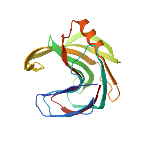
Crystallographic analysis shows substrate binding at the -3 to +1 active-site subsites and at the surface of glycoside hydrolase family 11 endo-1,4-beta-xylanases.
Vandermarliere, E., Bourgois, T.M., Rombouts, S., Van Campenhout, S., Volckaert, G., Strelkov, S.V., Delcour, J.A., Rabijns, A., Courtin, C.M.(2008) Biochem J 410: 71-79
- PubMed: 17983355 Search on PubMed
- DOI: https://doi.org/10.1042/BJ20071128
- Primary Citation Related Structures:
2QZ2, 2QZ3, 2Z79 - PubMed Abstract:
GH 11 (glycoside hydrolase family 11) xylanases are predominant enzymes in the hydrolysis of heteroxylan, an abundant structural polysaccharide in the plant cell wall. To gain more insight into the protein-ligand interactions of the glycone as well as the aglycone subsites of these enzymes, catalytically incompetent mutants of the Bacillus subtilis and Aspergillus niger xylanases were crystallized, soaked with xylo-oligosaccharides and subjected to X-ray analysis. For both xylanases, there was clear density for xylose residues in the -1 and -2 subsites. In addition, for the B. subtilis xylanase, there was also density for xylose residues in the -3 and +1 subsite showing the spanning of the -1/+1 subsites. These results, together with the observation that some residues in the aglycone subsites clearly adopt a different conformation upon substrate binding, allowed us to identify the residues important for substrate binding in the aglycone subsites. In addition to substrate binding in the active site of the enzymes, the existence of an unproductive second ligand-binding site located on the surface of both the B. subtilis and A. niger xylanases was observed. This extra binding site may have a function similar to the separate carbohydrate-binding modules of other glycoside hydrolase families.
- Laboratory for Biocrystallography, Department of Pharmaceutical Sciences, Katholieke Universiteit Leuven, Herestraat 49, O&N II, bus 822, 3000 Leuven, Belgium. Elien.Vandermarliere@pharm.kuleuven.be
Organizational Affiliation: