


Crystal structure of I-set domain of human Myosin Binding ProteinC
Kishishita, S., Ohsawa, N., Murayama, K., Terada, T., Chen, L., Liu, Z., Shirouzu, M., Wang, B., Yokoyama, S.To be published.
Experimental Data Snapshot
wwPDB Validation 3D Report Full Report

Entity ID: 1 | |||||
|---|---|---|---|---|---|
| Molecule | Chains | Sequence Length | Organism | Details | Image |
| Myosin-binding protein C, slow-type | 100 | Homo sapiens | Mutation(s): 0 | 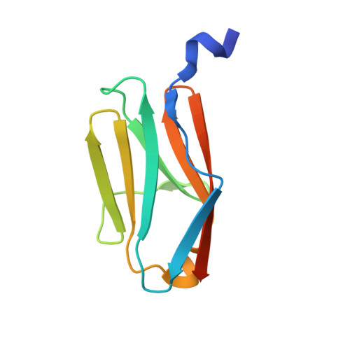 | |
UniProt & NIH Common Fund Data Resources | |||||
PHAROS: Q00872 GTEx: ENSG00000196091 | |||||
Entity Groups | |||||
| Sequence Clusters | 30% Identity50% Identity70% Identity90% Identity95% Identity100% Identity | ||||
| UniProt Group | Q00872 | ||||
Sequence AnnotationsExpand | |||||
Reference Sequence | |||||
| Length ( Å ) | Angle ( ˚ ) |
|---|---|
| a = 55.199 | α = 90 |
| b = 55.199 | β = 90 |
| c = 70.134 | γ = 90 |
| Software Name | Purpose |
|---|---|
| CNS | refinement |
| HKL-2000 | data reduction |
| HKL-2000 | data scaling |
| SOLVE | phasing |


