2YVL

Crystal structure of tRNA (m1A58) methyltransferase TrmI from Aquifex aeolicus


Experimental Data Snapshot

  • Method: X-RAY DIFFRACTION
  • Resolution: 2.20 Å
  • R-Value Free: 0.230 
  • R-Value Work: 0.194 
  • R-Value Observed: 0.196 

wwPDB Validation   3D Report Full Report


Ligand Structure Quality Assessment 


This is version 1.3 of the entry. See complete history


Literature

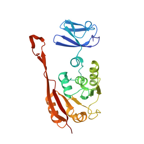
Crystal structure of tRNA m(1)A58 methyltransferase TrmI from Aquifex aeolicus in complex with S-adenosyl-L-methionine.

Kuratani, M.Yanagisawa, T.Ishii, R.Matsuno, M.Si, S.Y.Katsura, K.Ushikoshi-Nakayama, R.Shibata, R.Shirouzu, M.Bessho, Y.Yokoyama, S.

(2014) J Struct Funct Genomics 15: 173-180

  • DOI: https://doi.org/10.1007/s10969-014-9183-0
  • Primary Citation of Related Structures:  
    2YVL

  • PubMed Abstract: 

    The N (1)-methyladenosine residue at position 58 of tRNA is found in the three domains of life, and contributes to the stability of the three-dimensional L-shaped tRNA structure. In thermophilic bacteria, this modification is important for thermal adaptation, and is catalyzed by the tRNA m(1)A58 methyltransferase TrmI, using S-adenosyl-L-methionine (AdoMet) as the methyl donor. We present the 2.2 Å crystal structure of TrmI from the extremely thermophilic bacterium Aquifex aeolicus, in complex with AdoMet. There are four molecules per asymmetric unit, and they form a tetramer. Based on a comparison of the AdoMet binding mode of A. aeolicus TrmI to those of the Thermus thermophilus and Pyrococcus abyssi TrmIs, we discuss their similarities and differences. Although the binding modes to the N6 amino group of the adenine moiety of AdoMet are similar, using the side chains of acidic residues as well as hydrogen bonds, the positions of the amino acid residues involved in binding are diverse among the TrmIs from A. aeolicus, T. thermophilus, and P. abyssi.


  • Organizational Affiliation

    RIKEN Genomic Sciences Center, 1-7-22 Suehiro-cho, Tsurumi-ku, Yokohama, 230-0045, Japan.


Macromolecules
Find similar proteins by:  (by identity cutoff)  |  3D Structure
Entity ID: 1
MoleculeChains Sequence LengthOrganismDetailsImage
Hypothetical protein
A, B, C, D
248Aquifex aeolicus VF5Mutation(s): 0 
Gene Names: aq_311
EC: 2.1.1.36
UniProt
Find proteins for O66653 (Aquifex aeolicus (strain VF5))
Explore O66653 
Go to UniProtKB:  O66653
Entity Groups  
Sequence Clusters30% Identity50% Identity70% Identity90% Identity95% Identity100% Identity
UniProt GroupO66653
Sequence Annotations
Expand
  • Reference Sequence
Experimental Data & Validation

Experimental Data

  • Method: X-RAY DIFFRACTION
  • Resolution: 2.20 Å
  • R-Value Free: 0.230 
  • R-Value Work: 0.194 
  • R-Value Observed: 0.196 
  • Space Group: P 21 21 21
Unit Cell:
Length ( Å )Angle ( ˚ )
a = 69.826α = 90
b = 97.237β = 90
c = 212.687γ = 90
Software Package:
Software NamePurpose
REFMACrefinement
ADSCdata collection
HKL-2000data reduction
SCALEPACKdata scaling
MOLREPphasing

Structure Validation

View Full Validation Report


Ligand Structure Quality Assessment 


Entry History 

Deposition Data

Revision History  (Full details and data files)

  • Version 1.0: 2008-04-01
    Type: Initial release
  • Version 1.1: 2011-07-13
    Changes: Source and taxonomy, Version format compliance
  • Version 1.2: 2023-04-26
    Changes: Database references, Derived calculations
  • Version 1.3: 2023-11-08
    Changes: Data collection, Refinement description