2YVH

Crystal structure of the operator-binding form of the multi-drug binding transcriptional repressor CgmR


Experimental Data Snapshot

  • Method: X-RAY DIFFRACTION
  • Resolution: 2.50 Å
  • R-Value Free: 0.259 
  • R-Value Work: 0.220 
  • R-Value Observed: 0.222 

wwPDB Validation   3D Report Full Report


This is version 1.2 of the entry. See complete history


Literature

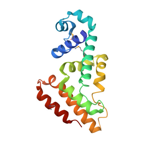
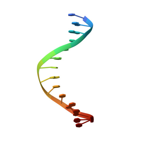
Crystal Structures of the Multidrug Binding Repressor Corynebacteriumglutamicum CgmR in Complex with Inducers and with an Operator

Itou, H.Watanabe, N.Yao, M.Shirakihara, Y.Tanaka, I.

(2010) J Mol Biol 403: 174-184

  • DOI: https://doi.org/10.1016/j.jmb.2010.07.042
  • Primary Citation of Related Structures:  
    2YVE, 2YVH, 2ZOZ

  • PubMed Abstract: 

    CgmR (CGL2612) from Corynebacterium glutamicum is a multidrug-resistance-related transcription factor belonging to the TetR family, which is a protein family of widespread bacterial transcription factors typically involved in environmental response. Here, we report the crystal structures of CgmR homodimeric repressor in complex with two distinct inducers (1.95 and 1.4 Å resolution) and with an operator (2.5 Å resolution). The CgmR-operator complex showed that two CgmR dimers bound to the operator, and each half-site of the palindromic operator was asymmetrically recognized by two DNA-binding domains from different dimers on the opposite sides of the DNA. The inducer complexes demonstrated that both bound inducers act as a wedge to alter the operator-binding conformation of the repressor by steric inhibition. As steric hindrance is used, various drugs should act as inducers if they have sufficient volume for the conformation change and if their bindings sufficiently reduce free energy. The comparative structural study of CgmR free protein, in complex with operator, and with inducers, implies the other mechanism that might contribute to multidrug response of the repressor.


  • Organizational Affiliation

    Structural Biology Center, National Institute of Genetics, Yata 1111, Mishima, Shizuoka 411-8540, Japan. hitou@lab.nig.ac.jp


Macromolecules

Find similar proteins by:  (by identity cutoff)  |  3D Structure
Entity ID: 1
MoleculeChains Sequence LengthOrganismDetailsImage
Transcriptional regulator
A, B, C, D
177Corynebacterium glutamicum ATCC 13032Mutation(s): 0 
Gene Names: cgl2612
UniProt
Find proteins for Q8NMG3 (Corynebacterium glutamicum (strain ATCC 13032 / DSM 20300 / BCRC 11384 / JCM 1318 / LMG 3730 / NCIMB 10025))
Explore Q8NMG3 
Go to UniProtKB:  Q8NMG3
Entity Groups  
Sequence Clusters30% Identity50% Identity70% Identity90% Identity95% Identity100% Identity
UniProt GroupQ8NMG3
Sequence Annotations
Expand
  • Reference Sequence

Find similar nucleic acids by:  Sequence   |   3D Structure  

Entity ID: 2
MoleculeChains LengthOrganismImage
5'-D(*DTP*DAP*DAP*DCP*DTP*DGP*DTP*DAP*DCP*DCP*DGP*DAP*DCP*DC)-3'
E, G
14N/A
Sequence Annotations
Expand
  • Reference Sequence

Find similar nucleic acids by:  Sequence   |   3D Structure  

Entity ID: 3
MoleculeChains LengthOrganismImage
5'-D(*DGP*DGP*DTP*DCP*DGP*DGP*DTP*DAP*DCP*DAP*DGP*DTP*DTP*DA)-3'
F, H
14N/A
Sequence Annotations
Expand
  • Reference Sequence
Small Molecules
Modified Residues  1 Unique
IDChains TypeFormula2D DiagramParent
MSE
Query on MSE
A, B, C, D
L-PEPTIDE LINKINGC5 H11 N O2 SeMET
Experimental Data & Validation

Experimental Data

  • Method: X-RAY DIFFRACTION
  • Resolution: 2.50 Å
  • R-Value Free: 0.259 
  • R-Value Work: 0.220 
  • R-Value Observed: 0.222 
  • Space Group: P 1
Unit Cell:
Length ( Å )Angle ( ˚ )
a = 39.011α = 108.98
b = 80.095β = 102.25
c = 87.056γ = 96.14
Software Package:
Software NamePurpose
REFMACrefinement
ADSCdata collection
HKL-2000data reduction
HKL-2000data scaling
SOLVEphasing

Structure Validation

View Full Validation Report


Entry History 

Deposition Data

Revision History  (Full details and data files)

  • Version 1.0: 2008-04-15
    Type: Initial release
  • Version 1.1: 2011-07-13
    Changes: Version format compliance
  • Version 1.2: 2017-01-11
    Changes: Other