2YV6 | pdb_00002yv6

Crystal structure of human Bcl-2 family protein Bak


Experimental Data Snapshot

  • Method: X-RAY DIFFRACTION
  • Resolution: 2.50 Å
  • R-Value Free: 
    0.248 (Depositor), 0.245 (DCC) 
  • R-Value Work: 
    0.214 (Depositor), 0.215 (DCC) 
  • R-Value Observed: 
    0.214 (Depositor) 

wwPDB Validation 3D Report Full Report

Validation slider image for 2YV6

This is version 1.3 of the entry. See complete history

Literature

Novel dimerization mode of the human Bcl-2 family protein Bak, a mitochondrial apoptosis regulator.

Wang, H.Takemoto, C.Akasaka, R.Uchikubo-Kamo, T.Kishishita, S.Murayama, K.Terada, T.Chen, L.Liu, Z.J.Wang, B.C.Sugano, S.Tanaka, A.Inoue, M.Kigawa, T.Shirouzu, M.Yokoyama, S.

(2009) J Struct Biol 166: 32-37

  • DOI: https://doi.org/10.1016/j.jsb.2008.12.003
  • Primary Citation Related Structures: 
    2YV6

  • PubMed Abstract: 

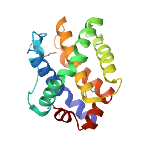
    Interactions of Bcl-2 family proteins play a regulatory role in mitochondrial apoptosis. The pro-apoptotic protein Bak resides in the outer mitochondrial membrane, and the formation of Bak homo- or heterodimers is involved in the regulation of apoptosis. The previously reported structure of the human Bak protein (residues Glu16-Gly186) revealed that a zinc ion was coordinated with two pairs of Asp160 and His164 residues from the symmetry-related molecules. This zinc-dependent homodimer was regarded as an anti-apoptotic dimer. In the present study, we determined the crystal structure of the human Bak residues Ser23-Asn185 at 2.5A, and found a distinct type of homodimerization through Cys166 disulfide bridging between the symmetry-related molecules. In the two modes of homodimerization, the molecular interfaces are completely different. In the membrane-targeted model of the S-S bridged dimer, the BH3 motifs are too close to the membrane to interact directly with the anti-apoptotic relatives, such as Bcl-x(L). Therefore, the Bak dimer structure reported here may represent a pro-apoptotic mode under oxidized conditions.


  • Organizational Affiliation
    • Systems and Structural Biology Center, Yokohama Institute, RIKEN, 1-7-22 Suehiro-cho, Tsurumi-ku, Yokohama 230-0045, Japan.

Macromolecule Content 

  • Total Structure Weight: 18.74 kDa 
  • Atom Count: 1,300 
  • Modeled Residue Count: 159 
  • Deposited Residue Count: 163 
  • Unique protein chains: 1

Macromolecules

Find similar proteins by:|  3D Structure
Entity ID: 1
MoleculeChains  Sequence LengthOrganismDetailsImage
Bcl-2 homologous antagonist/killer163Homo sapiensMutation(s): 0 
UniProt & NIH Common Fund Data Resources
Find proteins for Q16611 (Homo sapiens)
Explore Q16611 
Go to UniProtKB:  Q16611
PHAROS:  Q16611
GTEx:  ENSG00000030110 
Entity Groups
Sequence Clusters30% Identity50% Identity70% Identity90% Identity95% Identity100% Identity
UniProt GroupQ16611
Sequence Annotations
Expand
Reference Sequence

Small Molecules

Ligands 1 Unique
IDChains Name / Formula / InChI Key2D Diagram3D Interactions
SO4

Query on SO4


Download:Ideal Coordinates CCD File
B [auth A]SULFATE ION
O4 S
QAOWNCQODCNURD-UHFFFAOYSA-L
Modified Residues  1 Unique
IDChains TypeFormula2D DiagramParent
MSE
Query on MSE
A
L-PEPTIDE LINKINGC5 H11 N O2 SeMET

Experimental Data & Validation

Experimental Data

  • Method: X-RAY DIFFRACTION
  • Resolution: 2.50 Å
  • R-Value Free:  0.248 (Depositor), 0.245 (DCC) 
  • R-Value Work:  0.214 (Depositor), 0.215 (DCC) 
  • R-Value Observed: 0.214 (Depositor) 
Space Group: P 65 2 2
Unit Cell:
Length ( Å )Angle ( ˚ )
a = 61.824α = 90
b = 61.824β = 90
c = 137.482γ = 120
Software Package:
Software NamePurpose
CNSrefinement
HKL-2000data collection
HKL-2000data reduction
HKL-2000data scaling
SOLVEphasing

Structure Validation

View Full Validation Report


Entry History 

Revision History  (Full details and data files)

  • Version 1.0: 2008-04-15
    Type: Initial release
  • Version 1.1: 2008-10-08
    Changes: Version format compliance
  • Version 1.2: 2011-07-13
    Changes: Version format compliance
  • Version 1.3: 2024-10-30
    Changes: Data collection, Database references, Derived calculations, Structure summary