The Role of Protein Denaturation Energetics and Molecular Chaperones in the Aggregation and Mistargeting of Mutants Causing Primary Hyperoxaluria Type I
Mesa-Torres, N., Fabelo-Rosa, I., Riverol, D., Yunta, C., Albert, A., Salido, E., Pey, A.L.(2013) PLoS One 8: 71963
- PubMed: 24205397 Search on PubMedSearch on PubMed Central
- DOI: https://doi.org/10.1371/journal.pone.0071963
- Primary Citation Related Structures:
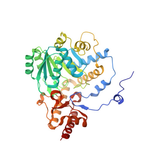
2YOB - PubMed Abstract:
Primary hyperoxaluria type I (PH1) is a conformational disease which result in the loss of alanine:glyoxylate aminotransferase (AGT) function. The study of AGT has important implications for protein folding and trafficking because PH1 mutants may cause protein aggregation and mitochondrial mistargeting. We herein describe a multidisciplinary study aimed to understand the molecular basis of protein aggregation and mistargeting in PH1 by studying twelve AGT variants. Expression studies in cell cultures reveal strong protein folding defects in PH1 causing mutants leading to enhanced aggregation, and in two cases, mitochondrial mistargeting. Immunoprecipitation studies in a cell-free system reveal that most mutants enhance the interactions with Hsc70 chaperones along their folding process, while in vitro binding experiments show no changes in the interaction of folded AGT dimers with the peroxisomal receptor Pex5p. Thermal denaturation studies by calorimetry support that PH1 causing mutants often kinetically destabilize the folded apo-protein through significant changes in the denaturation free energy barrier, whereas coenzyme binding overcomes this destabilization. Modeling of the mutations on a 1.9 Å crystal structure suggests that PH1 causing mutants perturb locally the native structure. Our work support that a misbalance between denaturation energetics and interactions with chaperones underlie aggregation and mistargeting in PH1, suggesting that native state stabilizers and protein homeostasis modulators are potential drugs to restore the complex and delicate balance of AGT protein homeostasis in PH1.
- Department of Physical Chemistry, Faculty of Sciences, University of Granada, Granada, Spain.
Organizational Affiliation: