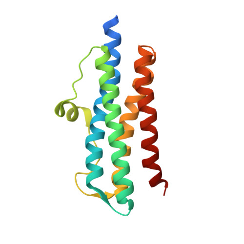
2YJJ

Structure of Dps from MICROBACTERIUM ARBORESCENS in the low iron form


Experimental Data Snapshot

  • Method: X-RAY DIFFRACTION
  • Resolution: 2.05 Å
  • R-Value Free: 0.226 
  • R-Value Work: 0.171 
  • R-Value Observed: 0.174 

wwPDB Validation   3D Report Full Report


This is version 1.2 of the entry. See complete history


Literature

Structure and Mechanism of Iron Translocation by a Dps Protein from Microbacterium Arborescens.

Pesek, J.Buecheler, R.Albrecht, R.Boland, W.Zeth, K.

(2011) J Biol Chem 286: 34872

  • DOI: https://doi.org/10.1074/jbc.M111.246108
  • Primary Citation of Related Structures:  
    2YJJ, 2YJK

  • PubMed Abstract: 

    Dps (DNA protection during starvation) enzymes are a major class of dodecameric proteins that bacteria use to detoxify their cytosol through the uptake of reactive iron species. In the stationary growth phase of bacteria, Dps enzymes are primarily used to protect DNA by biocrystallization. To characterize the wild type Dps protein from Microbacterium arborescens that displays additional catalytic functions (amide hydrolysis and synthesis), we determined the crystal structure to a resolution of 2.05 Å at low iron content. The structure shows a single iron at the ferroxidase center coordinated by an oxo atom, one water molecule, and three ligating residues. An iron-enriched protein structure was obtained at 2 Å and shows the stepwise uptake of two hexahydrated iron atoms moving along channels at the 3-fold axis before a restriction site inside the channels requires removal of the hydration sphere. Supporting biochemical data provide insight into the regulation of this acylamino acid hydrolase. Moreover, the peroxidase activity of the protein was determined. The influence of iron and siderophores on the expression of acylamino acid hydrolase was monitored during several stages of cell growth. Altogether our data provide an interesting view of an unusual Dps-like enzyme evolutionarily located apart from the large Dps sequence clusters.


  • Organizational Affiliation

    Department of Bioorganic Chemistry, Max Planck Institute for Chemical Ecology, Hans-Knöll-Strasse 8, D-07745 Jena, Germany.


Macromolecules
Find similar proteins by:  (by identity cutoff)  |  3D Structure
Entity ID: 1
MoleculeChains Sequence LengthOrganismDetailsImage
AFP
A, B, C, D, E
A, B, C, D, E, F, G, H, I, J, K, L
161Microbacterium arborescensMutation(s): 0 
UniProt
Find proteins for Q1X6M4 (Microbacterium arborescens)
Explore Q1X6M4 
Go to UniProtKB:  Q1X6M4
Entity Groups  
Sequence Clusters30% Identity50% Identity70% Identity90% Identity95% Identity100% Identity
UniProt GroupQ1X6M4
Sequence Annotations
Expand
  • Reference Sequence
Small Molecules
Ligands 2 Unique
IDChains Name / Formula / InChI Key2D Diagram3D Interactions
OFE
Query on OFE

Download Ideal Coordinates CCD File 
N [auth B]
O [auth C]
P [auth D]
Q [auth E]
R [auth F]
N [auth B],
O [auth C],
P [auth D],
Q [auth E],
R [auth F],
S [auth G],
T [auth H],
U [auth I],
V [auth J],
W [auth K]
Iron(II) oxide
Fe O
UQSXHKLRYXJYBZ-UHFFFAOYSA-N
FE
Query on FE

Download Ideal Coordinates CCD File 
M [auth A],
X [auth L]
FE (III) ION
Fe
VTLYFUHAOXGGBS-UHFFFAOYSA-N
Experimental Data & Validation

Experimental Data

  • Method: X-RAY DIFFRACTION
  • Resolution: 2.05 Å
  • R-Value Free: 0.226 
  • R-Value Work: 0.171 
  • R-Value Observed: 0.174 
  • Space Group: P 1 21 1
Unit Cell:
Length ( Å )Angle ( ˚ )
a = 87.53α = 90
b = 91.92β = 96.1
c = 128.45γ = 90
Software Package:
Software NamePurpose
REFMACrefinement
XDSdata reduction
XDSdata scaling
MOLREPphasing

Structure Validation

View Full Validation Report


Entry History 

Deposition Data

Revision History  (Full details and data files)

  • Version 1.0: 2011-06-29
    Type: Initial release
  • Version 1.1: 2011-10-12
    Changes: Database references, Version format compliance
  • Version 1.2: 2023-12-20
    Changes: Data collection, Database references, Derived calculations, Other, Refinement description