Functional Plasticity and Allosteric Regulation of Alpha-Ketoglutarate Decarboxylase in Central Mycobacterial Metabolism.
Wagner, T., Bellinzoni, M., Wehenkel, A.M., O'Hare, H.M., Alzari, P.M.(2011) Chem Biol 18: 1011
- PubMed: 21867916 Search on PubMed
- DOI: https://doi.org/10.1016/j.chembiol.2011.06.004
- Primary Citation Related Structures:
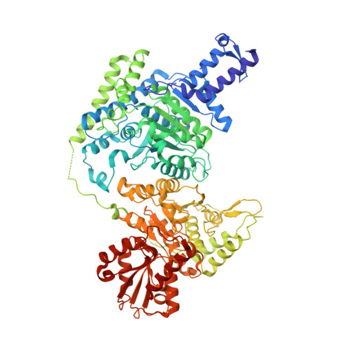
2XT6, 2XTA, 2Y0P, 2YIC, 2YID - PubMed Abstract:
The α-ketoglutarate dehydrogenase (KDH) complex is a major regulatory point of aerobic energy metabolism. Mycobacterium tuberculosis was reported to lack KDH activity, and the putative KDH E1o component, α-ketoglutarate decarboxylase (KGD), was instead assigned as a decarboxylase or carboligase. Here, we show that this protein does in fact sustain KDH activity, as well as the additional two reactions, and these multifunctional properties are shared by the Escherichia coli homolog, SucA. We also show that the mycobacterial enzyme is finely regulated by an additional acyltransferase-like domain and by the action of acetyl-CoA, a powerful allosteric activator able to enhance the concerted protein motions observed during catalysis. Our results uncover the functional plasticity of a crucial node in bacterial metabolism, which may be important for M. tuberculosis during host infection.
- Institut Pasteur, Unité de Biochimie Structurale (CNRS URA 2185), 25 rue du Dr. Roux, 75724 Paris, France.
Organizational Affiliation: