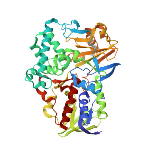
2YG5

Structure-based redesign of cofactor binding in Putrescine Oxidase: A394C mutant


Experimental Data Snapshot

  • Method: X-RAY DIFFRACTION
  • Resolution: 1.90 Å
  • R-Value Free: 0.242 
  • R-Value Work: 0.198 
  • R-Value Observed: 0.200 

Starting Model: experimental
View more details

wwPDB Validation   3D Report Full Report


Ligand Structure Quality Assessment 


This is version 1.2 of the entry. See complete history


Literature

Structure-Based Redesign of Cofactor Binding in Putrescine Oxidase.

Kopacz, M.M.Rovida, S.Van Duijn, E.Fraaije, M.W.Mattevi, A.

(2011) Biochemistry 50: 4209

  • DOI: https://doi.org/10.1021/bi200372u
  • Primary Citation of Related Structures:  
    2YG3, 2YG4, 2YG5, 2YG6, 2YG7

  • PubMed Abstract: 

    Putrescine oxidase (PuO) from Rhodococcus erythropolis is a soluble homodimeric flavoprotein, which oxidizes small aliphatic diamines. In this study, we report the crystal structures and cofactor binding properties of wild-type and mutant enzymes. From a structural viewpoint, PuO closely resembles the sequence-related human monoamine oxidases A and B. This similarity is striking in the flavin-binding site even if PuO does not covalently bind the cofactor as do the monoamine oxidases. A remarkable conserved feature is the cis peptide conformation of the Tyr residue whose conformation is important for substrate recognition in the active site cavity. The structure of PuO in complex with the reaction product reveals that Glu324 is crucial in recognizing the terminal amino group of the diamine substrate and explains the narrow substrate specificity of the enzyme. The structural analysis also provides clues for identification of residues that are responsible for the competitive binding of ADP versus FAD (~50% of wild-type PuO monomers isolated are occupied by ADP instead of FAD). By replacing Pro15, which is part of the dinucleotide-binding domain, enzyme preparations were obtained that are almost 100% in the FAD-bound form. Furthermore, mutants have been designed and prepared that form a covalent 8α-S-cysteinyl-FAD linkage. These data provide new insights into the molecular basis for substrate recognition in amine oxidases and demonstrate that engineering of flavoenzymes to introduce covalent linkage with the cofactor is a possible route to develop more stable protein molecules, better suited for biocatalytic purposes.


  • Organizational Affiliation

    Laboratory of Biochemistry, Groningen Biomolecular Sciences and Biotechnology Institute, University of Groningen, Nijenborgh 4, 9747 AG Groningen, The Netherlands.


Macromolecules
Find similar proteins by:  (by identity cutoff)  |  3D Structure
Entity ID: 1
MoleculeChains Sequence LengthOrganismDetailsImage
PUTRESCINE OXIDASE453Rhodococcus erythropolisMutation(s): 1 
EC: 1.4.3.10
UniProt
Find proteins for B0F9F6 (Rhodococcus erythropolis)
Explore B0F9F6 
Go to UniProtKB:  B0F9F6
Entity Groups  
Sequence Clusters30% Identity50% Identity70% Identity90% Identity95% Identity100% Identity
UniProt GroupB0F9F6
Sequence Annotations
Expand
  • Reference Sequence
Small Molecules
Ligands 3 Unique
IDChains Name / Formula / InChI Key2D Diagram3D Interactions
FAD
Query on FAD

Download Ideal Coordinates CCD File 
B [auth A]FLAVIN-ADENINE DINUCLEOTIDE
C27 H33 N9 O15 P2
VWWQXMAJTJZDQX-UYBVJOGSSA-N
SO4
Query on SO4

Download Ideal Coordinates CCD File 
C [auth A],
D [auth A],
G [auth A]
SULFATE ION
O4 S
QAOWNCQODCNURD-UHFFFAOYSA-L
GOL
Query on GOL

Download Ideal Coordinates CCD File 
E [auth A],
F [auth A],
H [auth A]
GLYCEROL
C3 H8 O3
PEDCQBHIVMGVHV-UHFFFAOYSA-N
Experimental Data & Validation

Experimental Data

  • Method: X-RAY DIFFRACTION
  • Resolution: 1.90 Å
  • R-Value Free: 0.242 
  • R-Value Work: 0.198 
  • R-Value Observed: 0.200 
  • Space Group: P 41 2 2
Unit Cell:
Length ( Å )Angle ( ˚ )
a = 102.003α = 90
b = 102.003β = 90
c = 130.361γ = 90
Software Package:
Software NamePurpose
REFMACrefinement
MOSFLMdata reduction
SCALAdata scaling
CCP4phasing

Structure Validation

View Full Validation Report


Ligand Structure Quality Assessment 


Entry History 

Deposition Data

Revision History  (Full details and data files)

  • Version 1.0: 2011-05-18
    Type: Initial release
  • Version 1.1: 2011-07-13
    Changes: Version format compliance
  • Version 1.2: 2023-12-20
    Changes: Data collection, Database references, Derived calculations, Other, Refinement description