Crystal Structures of the Endoplasmic Reticulum Aminopeptidase-1 (Erap1) Reveal the Molecular Basis for N-Terminal Peptide Trimming.
Kochan, G., Krojer, T., Harvey, D., Fischer, R., Chen, L., Vollmar, M., von Delft, F., Kavanagh, K.L., Brown, M.A., Bowness, P., Wordsworth, P., Kessler, B.M., Oppermann, U.(2011) Proc Natl Acad Sci U S A 108: 7745
- PubMed: 21508329 Search on PubMedSearch on PubMed Central
- DOI: https://doi.org/10.1073/pnas.1101262108
- Primary Citation Related Structures:
2YD0, 3QNF - PubMed Abstract:
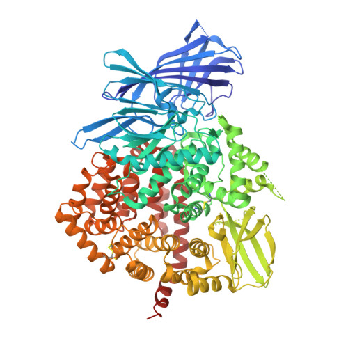
Endoplasmatic reticulum aminopeptidase 1 (ERAP1) is a multifunctional enzyme involved in trimming of peptides to an optimal length for presentation by major histocompatibility complex (MHC) class I molecules. Polymorphisms in ERAP1 have been associated with chronic inflammatory diseases, including ankylosing spondylitis (AS) and psoriasis, and subsequent in vitro enzyme studies suggest distinct catalytic properties of ERAP1 variants. To understand structure-activity relationships of this enzyme we determined crystal structures in open and closed states of human ERAP1, which provide the first snapshots along a catalytic path. ERAP1 is a zinc-metallopeptidase with typical H-E-X-X-H-(X)(18)-E zinc binding and G-A-M-E-N motifs characteristic for members of the gluzincin protease family. The structures reveal extensive domain movements, including an active site closure as well as three different open conformations, thus providing insights into the catalytic cycle. A K(528)R mutant strongly associated with AS in GWAS studies shows significantly altered peptide processing characteristics, which are possibly related to impaired interdomain interactions.
- Structural Genomics Consortium, University of Oxford, Old Road Campus, Roosevelt Drive, Headington OX3 7DQ, United Kingdom.
Organizational Affiliation: