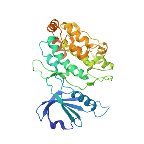
Structure of the Dimeric Autoinhibited Conformation of Dapk2, a Pro-Apoptotic Protein Kinase.
Patel, A.K., Yadav, R.P., Majava, V., Kursula, I., Kursula, P.(2011) J Mol Biology 409: 369
- PubMed: 21497605
- DOI: https://doi.org/10.1016/j.jmb.2011.03.065
- Primary Citation of Related Structures:
2YA9, 2YAA, 2YAB - PubMed Abstract:
The death-associated protein kinase (DAPK) family has been characterized as a group of pro-apoptotic serine/threonine kinases that share specific structural features in their catalytic kinase domain. Two of the DAPK family members, DAPK1 and DAPK2, are calmodulin-dependent protein kinases that are regulated by oligomerization, calmodulin binding, and autophosphorylation. In this study, we have determined the crystal and solution structures of murine DAPK2 in the presence of the autoinhibitory domain, with and without bound nucleotides in the active site. The crystal structure shows dimers of DAPK2 in a conformation that is not permissible for protein substrate binding. Two different conformations were seen in the active site upon the introduction of nucleotide ligands. The monomeric and dimeric forms of DAPK2 were further analyzed for solution structure, and the results indicate that the dimers of DAPK2 are indeed formed through the association of two apposed catalytic domains, as seen in the crystal structure. The structures can be further used to build a model for DAPK2 autophosphorylation and to compare with closely related kinases, of which especially DAPK1 is an actively studied drug target. Our structures also provide a model for both homodimerization and heterodimerization of the catalytic domain between members of the DAPK family. The fingerprint of the DAPK family, the basic loop, plays a central role in the dimerization of the kinase domain.
- Department of Biochemistry, University of Oulu, Finland.
Organizational Affiliation:


