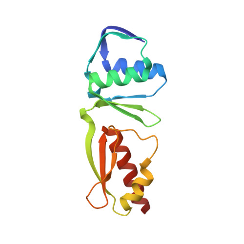
2Y9K

Three-dimensional model of Salmonella's needle complex at subnanometer resolution


Experimental Data Snapshot

  • Method: ELECTRON MICROSCOPY
  • Resolution: 8.30 Å
  • Aggregation State: PARTICLE 
  • Reconstruction Method: SINGLE PARTICLE 

wwPDB Validation   3D Report Full Report


This is version 1.4 of the entry. See complete history


Literature

Three-Dimensional Model of Salmonella'S Needle Complex at Subnanometer Resolution.

Schraidt, O.Marlovits, T.C.

(2011) Science 331: 1192

  • DOI: https://doi.org/10.1126/science.1199358
  • Primary Citation of Related Structures:  
    2Y9J, 2Y9K

  • PubMed Abstract: 

    Type III secretion systems (T3SSs) are essential virulence factors used by many Gram-negative bacteria to inject proteins that make eukaryotic host cells accessible to invasion. The T3SS core structure, the needle complex (NC), is a ~3.5 megadalton-sized, oligomeric, membrane-embedded complex. Analyzing cryo-electron microscopy images of top views of NCs or NC substructures from Salmonella typhimurium revealed a 24-fold symmetry for the inner rings and a 15-fold symmetry for the outer rings, giving an overall C3 symmetry. Local refinement and averaging showed the organization of the central core and allowed us to reconstruct a subnanometer composite structure of the NC, which together with confident docking of atomic structures reveal insights into its overall organization and structural requirements during assembly.


  • Organizational Affiliation

    Research Institute of Molecular Pathology, Dr. Bohr Gasse 7, A-1030 Vienna, Austria.


Macromolecules
Find similar proteins by:  (by identity cutoff)  |  3D Structure
Entity ID: 1
MoleculeChains Sequence LengthOrganismDetailsImage
PROTEIN INVG
A, B, C, D, E
A, B, C, D, E, F, G, H, I, J, K, L, M, N, O
137Salmonella enterica subsp. enterica serovar TyphimuriumMutation(s): 0 
UniProt
Find proteins for P35672 (Salmonella typhimurium (strain LT2 / SGSC1412 / ATCC 700720))
Explore P35672 
Go to UniProtKB:  P35672
Entity Groups  
Sequence Clusters30% Identity50% Identity70% Identity90% Identity95% Identity100% Identity
UniProt GroupP35672
Sequence Annotations
Expand
  • Reference Sequence
Experimental Data & Validation

Experimental Data

  • Method: ELECTRON MICROSCOPY
  • Resolution: 8.30 Å
  • Aggregation State: PARTICLE 
  • Reconstruction Method: SINGLE PARTICLE 
EM Software:
TaskSoftware PackageVersion
RECONSTRUCTIONIMAGIC

Structure Validation

View Full Validation Report


Entry History 

Deposition Data

Revision History  (Full details and data files)

  • Version 1.0: 2011-03-23
    Type: Initial release
  • Version 1.1: 2013-03-20
    Changes: Other, Refinement description, Version format compliance
  • Version 1.2: 2017-04-19
    Changes: Other
  • Version 1.3: 2017-08-30
    Changes: Data collection, Refinement description
  • Version 1.4: 2019-10-23
    Changes: Data collection, Other