Structural and Computational Investigations of Vim- 7: Insights Into the Substrate Specificity of Vim Metallo-Beta-Lactamases
Saradhi, P., Leiros, H.-K.S., Ahmad, R., Spencer, J., Leiros, I., Walsh, T.R., Sundsfjord, A., Samuelsen, O.(2011) J Mol Biology 411: 174
- PubMed: 21645522 Search on PubMed
- DOI: https://doi.org/10.1016/j.jmb.2011.05.035
- Primary Citation Related Structures:
2Y87, 2Y8A, 2Y8B - PubMed Abstract:
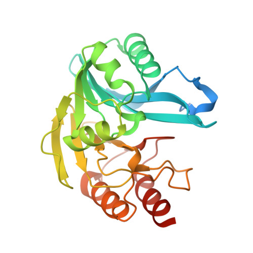
The presence of metallo-β-lactamases (MBLs) in many clinically important human bacterial pathogens limits treatment options, as these enzymes efficiently hydrolyze nearly all β-lactam antibiotics. VIM enzymes are among the most widely distributed MBLs, but many of the individual VIM subtypes remain poorly characterized. Pseudomonas aeruginosa VIM-7 is the most divergent among VIM-type MBLs in terms of amino acid sequence. Here we present crystal structures of VIM-7 as the native enzyme, with Cys221 oxidized (VIM-7-Ox), and with a sulfur atom bridging the two active-site zinc ions (VIM-7-S). Comparison with VIM-2 and VIM-4 structures suggests an explanation for the reduced catalytic efficiency of VIM-7 against cephalosporins with a positively charged cyclic substituent at the C3 position (e.g., ceftazidime). Kinetic variations are attributed to substitutions in residues 60-66 (that form a loop adjacent to the active site previously implicated in substrate binding) and to the disruption of two hydrogen-bonding clusters through substitutions at positions 218 and 224. Furthermore, the less negatively charged surface of VIM-7 (compared to VIM-2) may also contribute to the reduced hydrolytic efficiency. Docking of the cephalosporins ceftazidime and cefotaxime into the VIM-2 and VIM-7 structures reveals that amino acid substitutions may cause the mode of substrate binding to differ between the two enzymes. Our structures thus provide new insights into the variation in substrate specificity that is evident across this family of clinically important enzymes.
- Research Group for Host-Microbe Interactions, Department of Medical Biology, Faculty of Health Sciences, University of Tromsø, Tromsø, Norway.
Organizational Affiliation: