2Y5E

BARLEY LIMIT DEXTRINASE IN COMPLEX WITH ALPHA-CYCLODEXTRIN


Experimental Data Snapshot

  • Method: X-RAY DIFFRACTION
  • Resolution: 2.49 Å
  • R-Value Free: 0.226 
  • R-Value Work: 0.175 
  • R-Value Observed: 0.175 

wwPDB Validation   3D Report Full Report


This is version 2.0 of the entry. See complete history


Literature

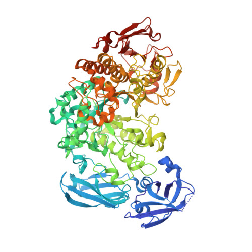
Crystal Structure of an Essential Enzyme in Seed Starch Degradation: Barley Limit Dextrinase in Complex with Cyclodextrins.

Vester-Christensen, M.B.Abou Hachem, M.Svensson, B.Henriksen, A.

(2010) J Mol Biol 403: 739

  • DOI: 10.1016/j.jmb.2010.09.031
  • Primary Citation of Related Structures:  
    2Y4S, 2Y5E

  • PubMed Abstract: 
  • Barley limit dextrinase [Hordeum vulgare limit dextrinase (HvLD)] catalyzes the hydrolysis of α-1,6 glucosidic linkages in limit dextrins. This activity plays a role in starch degradation during germination and presumably in starch biosynthesis during grain filling ...

    Barley limit dextrinase [Hordeum vulgare limit dextrinase (HvLD)] catalyzes the hydrolysis of α-1,6 glucosidic linkages in limit dextrins. This activity plays a role in starch degradation during germination and presumably in starch biosynthesis during grain filling. The crystal structures of HvLD in complex with the competitive inhibitors α-cyclodextrin (CD) and β-CD are solved and refined to 2.5 Å and 2.1 Å, respectively, and are the first structures of a limit dextrinase. HvLD belongs to glycoside hydrolase 13 family and is composed of four domains: an immunoglobulin-like N-terminal eight-stranded β-sandwich domain, a six-stranded β-sandwich domain belonging to the carbohydrate binding module 48 family, a catalytic (β/α)(8)-like barrel domain that lacks α-helix 5, and a C-terminal eight-stranded β-sandwich domain of unknown function. The CDs are bound at the active site occupying carbohydrate binding subsites +1 and +2. A glycerol and three water molecules mimic a glucose residue at subsite -1, thereby identifying residues involved in catalysis. The bulky Met440, a unique residue at its position among α-1,6 acting enzymes, obstructs subsite -4. The steric hindrance observed is proposed to affect substrate specificity and to cause a low activity of HvLD towards amylopectin. An extended loop (Asp513-Asn520) between β5 and β6 of the catalytic domain also seems to influence substrate specificity and to give HvLD a higher affinity for α-CD than pullulanases. The crystal structures additionally provide new insight into cation sites and the concerted action of the battery of hydrolytic enzymes in starch degradation.


    Related Citations: 
    • Secretory Expression of Functional Barley Limit Dextrinase by Pichia Pastoris Using High Cell- Density Fermentation.
      Vester-Christensen, M.B., Hachem, M.A., Naested, H., Svensson, B.
      (2010) Protein Expr Purif 69: 112

    Organizational Affiliation

    Enzyme and Protein Chemistry, Department of Systems Biology, Søltofts Plads, Building 224, Technical University of Denmark, Kgs. Lyngby, Denmark.


Macromolecules
Find similar proteins by:  (by identity cutoff)  |  3D Structure
Entity ID: 1
MoleculeChainsSequence LengthOrganismDetailsImage
LIMIT DEXTRINASEA884Hordeum vulgareMutation(s): 3 
EC: 3.2.1.41
UniProt
Find proteins for O48541 (Hordeum vulgare subsp. vulgare)
Explore O48541 
Go to UniProtKB:  O48541
Entity Groups  
Sequence Clusters30% Identity50% Identity70% Identity90% Identity95% Identity100% Identity
UniProt GroupO48541
Protein Feature View
Expand
  • Reference Sequence
Oligosaccharides

Help

Entity ID: 2
MoleculeChainsChain Length2D DiagramGlycosylation3D Interactions
Cyclohexakis-(1-4)-(alpha-D-glucopyranose)B 6N/A Oligosaccharides Interaction
Glycosylation Resources
GlyTouCan:  G22307HL
GlyCosmos:  G22307HL
Biologically Interesting Molecules (External Reference) 1 Unique
Entity ID: 2
IDChainsNameType/Class2D Diagram3D Interactions
PRD_900015
Query on PRD_900015
Balpha-cyclodextrinOligosaccharide / Drug delivery Ligand Interaction
Experimental Data & Validation

Experimental Data

  • Method: X-RAY DIFFRACTION
  • Resolution: 2.49 Å
  • R-Value Free: 0.226 
  • R-Value Work: 0.175 
  • R-Value Observed: 0.175 
  • Space Group: C 1 2 1
Unit Cell:
Length ( Å )Angle ( ˚ )
a = 174.84α = 90
b = 85.61β = 96.83
c = 61.12γ = 90
Software Package:
Software NamePurpose
PHENIXrefinement
MOSFLMdata reduction
SCALAdata scaling
PHASERphasing

Structure Validation

View Full Validation Report


Entry History 

Deposition Data

Revision History  (Full details and data files)

  • Version 1.0: 2011-01-26
    Type: Initial release
  • Version 1.1: 2012-11-21
    Changes: Database references, Non-polymer description
  • Version 1.2: 2018-01-17
    Changes: Data collection
  • Version 2.0: 2020-07-29
    Type: Remediation
    Reason: Carbohydrate remediation
    Changes: Atomic model, Data collection, Derived calculations, Non-polymer description, Other, Structure summary