2Y4R

CRYSTAL STRUCTURE OF 4-AMINO-4-DEOXYCHORISMATE LYASE FROM PSEUDOMONAS AERUGINOSA


Experimental Data Snapshot

  • Method: X-RAY DIFFRACTION
  • Resolution: 1.75 Å
  • R-Value Free: 0.215 
  • R-Value Work: 0.167 
  • R-Value Observed: 0.169 

wwPDB Validation   3D Report Full Report


Ligand Structure Quality Assessment 


This is version 1.4 of the entry. See complete history


Literature

Pseudomonas Aeruginosa 4-Amino-4-Deoxychorismate Lyase: Spatial Conservation of an Active Site Tyrosine and Classification of Two Types of Enzyme.

O'Rourke, P.E.F.Eadsforth, T.C.Fyfe, P.K.Shepherd, S.M.Hunter, W.N.

(2011) PLoS One 6: 24158

  • DOI: https://doi.org/10.1371/journal.pone.0024158
  • Primary Citation of Related Structures:  
    2Y4R

  • PubMed Abstract: 

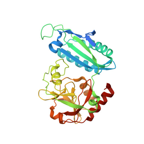
    4-Amino-4-deoxychorismate lyase (PabC) catalyzes the formation of 4-aminobenzoate, and release of pyruvate, during folate biosynthesis. This is an essential activity for the growth of gram-negative bacteria, including important pathogens such as Pseudomonas aeruginosa. A high-resolution (1.75 Å) crystal structure of PabC from P. aeruginosa has been determined, and sequence-structure comparisons with orthologous structures are reported. Residues around the pyridoxal 5'-phosphate cofactor are highly conserved adding support to aspects of a mechanism generic for enzymes carrying that cofactor. However, we suggest that PabC can be classified into two groups depending upon whether an active site and structurally conserved tyrosine is provided from the polypeptide that mainly forms an active site or from the partner subunit in the dimeric assembly. We considered that the conserved tyrosine might indicate a direct role in catalysis: that of providing a proton to reduce the olefin moiety of substrate as pyruvate is released. A threonine had previously been suggested to fulfill such a role prior to our observation of the structurally conserved tyrosine. We have been unable to elucidate an experimentally determined structure of PabC in complex with ligands to inform on mechanism and substrate specificity. Therefore we constructed a computational model of the catalytic intermediate docked into the enzyme active site. The model suggests that the conserved tyrosine helps to create a hydrophobic wall on one side of the active site that provides important interactions to bind the catalytic intermediate. However, this residue does not appear to participate in interactions with the C atom that undergoes an sp(2) to sp(3) conversion as pyruvate is produced. The model and our comparisons rather support the hypothesis that an active site threonine hydroxyl contributes a proton used in the reduction of the substrate methylene to pyruvate methyl in the final stage of the mechanism.


  • Organizational Affiliation

    Division of Biological Chemistry and Drug Discovery, College of Life Sciences, University of Dundee, Dundee, United Kingdom.


Macromolecules
Find similar proteins by:  (by identity cutoff)  |  3D Structure
Entity ID: 1
MoleculeChains Sequence LengthOrganismDetailsImage
4-AMINO-4-DEOXYCHORISMATE LYASE
A, B
292Pseudomonas aeruginosa PAO1Mutation(s): 0 
EC: 4.1.3.38
UniProt
Find proteins for Q9HZN6 (Pseudomonas aeruginosa (strain ATCC 15692 / DSM 22644 / CIP 104116 / JCM 14847 / LMG 12228 / 1C / PRS 101 / PAO1))
Explore Q9HZN6 
Go to UniProtKB:  Q9HZN6
Entity Groups  
Sequence Clusters30% Identity50% Identity70% Identity90% Identity95% Identity100% Identity
UniProt GroupQ9HZN6
Sequence Annotations
Expand
  • Reference Sequence
Small Molecules
Ligands 7 Unique
IDChains Name / Formula / InChI Key2D Diagram3D Interactions
PLP
Query on PLP

Download Ideal Coordinates CCD File 
C [auth A],
O [auth B]
PYRIDOXAL-5'-PHOSPHATE
C8 H10 N O6 P
NGVDGCNFYWLIFO-UHFFFAOYSA-N
PG4
Query on PG4

Download Ideal Coordinates CCD File 
D [auth A],
E [auth A],
F [auth A],
P [auth B],
Q [auth B]
TETRAETHYLENE GLYCOL
C8 H18 O5
UWHCKJMYHZGTIT-UHFFFAOYSA-N
PEG
Query on PEG

Download Ideal Coordinates CCD File 
L [auth A],
R [auth B],
U [auth B]
DI(HYDROXYETHYL)ETHER
C4 H10 O3
MTHSVFCYNBDYFN-UHFFFAOYSA-N
SO4
Query on SO4

Download Ideal Coordinates CCD File 
I [auth A],
S [auth B]
SULFATE ION
O4 S
QAOWNCQODCNURD-UHFFFAOYSA-L
GOL
Query on GOL

Download Ideal Coordinates CCD File 
M [auth A],
V [auth B]
GLYCEROL
C3 H8 O3
PEDCQBHIVMGVHV-UHFFFAOYSA-N
EDO
Query on EDO

Download Ideal Coordinates CCD File 
J [auth A],
K [auth A],
T [auth B]
1,2-ETHANEDIOL
C2 H6 O2
LYCAIKOWRPUZTN-UHFFFAOYSA-N
CL
Query on CL

Download Ideal Coordinates CCD File 
G [auth A],
H [auth A],
N [auth A]
CHLORIDE ION
Cl
VEXZGXHMUGYJMC-UHFFFAOYSA-M
Experimental Data & Validation

Experimental Data

  • Method: X-RAY DIFFRACTION
  • Resolution: 1.75 Å
  • R-Value Free: 0.215 
  • R-Value Work: 0.167 
  • R-Value Observed: 0.169 
  • Space Group: P 21 21 21
Unit Cell:
Length ( Å )Angle ( ˚ )
a = 40.813α = 90
b = 66.817β = 90
c = 202.688γ = 90
Software Package:
Software NamePurpose
REFMACrefinement
iMOSFLMdata reduction
SCALAdata scaling
MOLREPphasing

Structure Validation

View Full Validation Report


Ligand Structure Quality Assessment 


Entry History 

Deposition Data

Revision History  (Full details and data files)

  • Version 1.0: 2011-01-19
    Type: Initial release
  • Version 1.1: 2011-10-05
    Changes: Database references, Non-polymer description
  • Version 1.2: 2019-01-30
    Changes: Advisory, Data collection, Experimental preparation, Other
  • Version 1.3: 2019-03-06
    Changes: Data collection, Derived calculations, Experimental preparation
  • Version 1.4: 2023-12-20
    Changes: Advisory, Data collection, Database references, Derived calculations, Other, Refinement description