2XTJ | pdb_00002xtj

The crystal structure of PCSK9 in complex with 1D05 Fab


Experimental Data Snapshot

  • Method: X-RAY DIFFRACTION
  • Resolution: 2.70 Å
  • R-Value Free: 
    0.259 (Depositor), 0.259 (DCC) 
  • R-Value Work: 
    0.200 (Depositor), 0.199 (DCC) 
  • R-Value Observed: 
    0.203 (Depositor) 

Starting Model: experimental
View more details

wwPDB Validation 3D Report Full Report

Validation slider image for 2XTJ

This is version 1.5 of the entry. See complete history

Literature

A Pcsk9-Binding Antibody that Structurally Mimics the Egf(A) Domain of Ldl-Receptor Reduces Ldl Cholesterol in Vivo.

Ni, Y.G.Di Marco, S.Condra, J.H.Peterson, L.B.Wang, W.Wang, F.Pandit, S.Hammond, H.A.Rosa, R.Cummings, R.T.Wood, D.D.Liu, X.Bottomley, M.J.Shen, X.Cubbon, R.M.Wang, S.P.Johns, D.G.Volpari, C.Hamuro, L.Chin, J.Huang, L.Zhao, J.Z.Vitelli, S.Haytko, P.Wisniewski, D.Mitnaul, L.J.Sparrow, C.P.Hubbard, B.Carfi, A.Sitlani, A.

(2011) J Lipid Res 52: 78

  • DOI: https://doi.org/10.1194/jlr.M011445
  • Primary Citation Related Structures: 
    2XTJ

  • PubMed Abstract: 

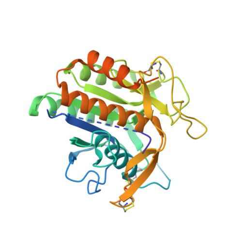
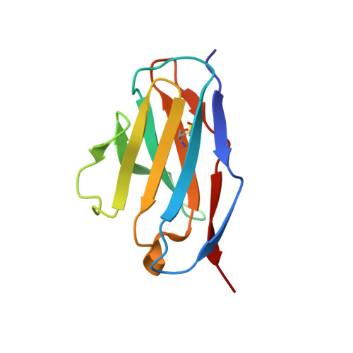
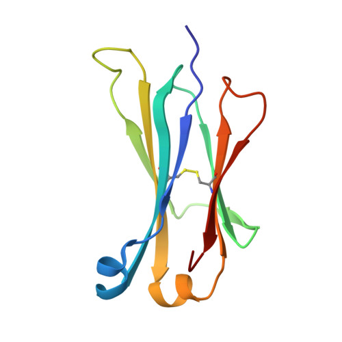
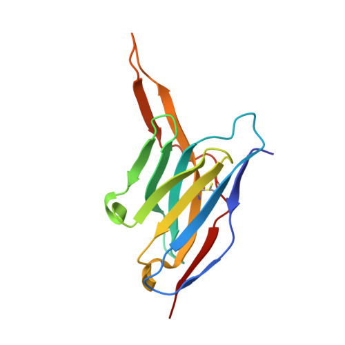
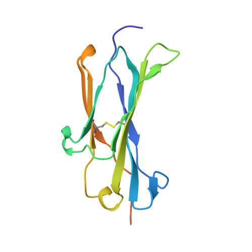
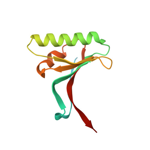
    Proprotein convertase subtilisin-like/kexin type 9 (PCSK9) regulates LDL cholesterol levels by inhibiting LDL receptor (LDLr)-mediated cellular LDL uptake. We have identified a fragment antigen-binding (Fab) 1D05 which binds PCSK9 with nanomolar affinity. The fully human antibody 1D05-IgG2 completely blocks the inhibitory effects of wild-type PCSK9 and two gain-of-function human PCSK9 mutants, S127R and D374Y. The crystal structure of 1D05-Fab bound to PCSK9 reveals that 1D05-Fab binds to an epitope on the PCSK9 catalytic domain which includes the entire LDLr EGF(A) binding site. Notably, the 1D05-Fab CDR-H3 and CDR-H2 loops structurally mimic the EGF(A) domain of LDLr. In a transgenic mouse model (CETP/LDLr-hemi), in which plasma lipid and PCSK9 profiles are comparable to those of humans, 1D05-IgG2 reduces plasma LDL cholesterol to 40% and raises hepatic LDLr protein levels approximately fivefold. Similarly, in healthy rhesus monkeys, 1D05-IgG2 effectively reduced LDL cholesterol 20%-50% for over 2 weeks, despite its relatively short terminal half-life (t(1/2) = 3.2 days). Importantly, the decrease in circulating LDL cholesterol corresponds closely to the reduction in free PCSK9 levels. Together these results clearly demonstrate that the LDL-lowering effect of the neutralizing anti-PCSK9 1D05-IgG2 antibody is mediated by reducing the amount of PCSK9 that can bind to the LDLr.


  • Organizational Affiliation
    • Department of Cardiovascular Diseases, Merck Research Laboratories, Rahway, New Jersey, USA. yan_ni@merck.com

Macromolecule Content 

  • Total Structure Weight: 96.43 kDa 
  • Atom Count: 6,096 
  • Modeled Residue Count: 782 
  • Deposited Residue Count: 894 
  • Unique protein chains: 6

Macromolecules

Find similar proteins by:|  3D Structure
Entity ID: 1
MoleculeChains  Sequence LengthOrganismDetailsImage
PROPROTEIN CONVERTASE SUBTILISIN/KEXIN TYPE 9312Homo sapiensMutation(s): 0 
EC: 3.4.21
UniProt & NIH Common Fund Data Resources
Find proteins for Q8NBP7 (Homo sapiens)
Explore Q8NBP7 
Go to UniProtKB:  Q8NBP7
PHAROS:  Q8NBP7
GTEx:  ENSG00000169174 
Entity Groups
Sequence Clusters30% Identity50% Identity70% Identity90% Identity95% Identity100% Identity
UniProt GroupQ8NBP7
Sequence Annotations
Expand
Reference Sequence
Find similar proteins by:|  3D Structure
Entity ID: 2
MoleculeChains  Sequence LengthOrganismDetailsImage
FAB FROM A HUMAN MONOCLONAL ANTIBODY, 1D05107Homo sapiensMutation(s): 0 
Find similar proteins by:|  3D Structure
Entity ID: 3
MoleculeChains  Sequence LengthOrganismDetailsImage
FAB FROM A HUMAN MONOCLONAL ANTIBODY, 1D05106Homo sapiensMutation(s): 0 
Find similar proteins by:|  3D Structure
Entity ID: 4
MoleculeChains  Sequence LengthOrganismDetailsImage
FAB FROM A HUMAN MONOCLONAL ANTIBODY, 1D05131Homo sapiensMutation(s): 0 
Find similar proteins by:|  3D Structure
Entity ID: 5
MoleculeChains  Sequence LengthOrganismDetailsImage
FAB FROM A HUMAN MONOCLONAL ANTIBODY, 1D05124Homo sapiensMutation(s): 0 
Find similar proteins by:|  3D Structure
Entity ID: 6
MoleculeChains  Sequence LengthOrganismDetailsImage
PROPROTEIN CONVERTASE SUBTILISIN-KEXIN TYPE 9F [auth P]114Homo sapiensMutation(s): 0 
EC: 3.4.21
UniProt & NIH Common Fund Data Resources
Find proteins for Q8NBP7 (Homo sapiens)
Explore Q8NBP7 
Go to UniProtKB:  Q8NBP7
PHAROS:  Q8NBP7
GTEx:  ENSG00000169174 
Entity Groups
Sequence Clusters30% Identity50% Identity70% Identity90% Identity95% Identity100% Identity
UniProt GroupQ8NBP7
Sequence Annotations
Expand
Reference Sequence

Small Molecules

Ligands 1 Unique
IDChains Name / Formula / InChI Key2D Diagram3D Interactions
CA

Query on CA


Download:Ideal Coordinates CCD File
G [auth A]CALCIUM ION
Ca
BHPQYMZQTOCNFJ-UHFFFAOYSA-N

Experimental Data & Validation

Experimental Data

  • Method: X-RAY DIFFRACTION
  • Resolution: 2.70 Å
  • R-Value Free:  0.259 (Depositor), 0.259 (DCC) 
  • R-Value Work:  0.200 (Depositor), 0.199 (DCC) 
  • R-Value Observed: 0.203 (Depositor) 
Space Group: P 21 21 21
Unit Cell:
Length ( Å )Angle ( ˚ )
a = 66.589α = 90
b = 67.834β = 90
c = 250.86γ = 90
Software Package:
Software NamePurpose
REFMACrefinement
MOSFLMdata reduction
SCALAdata scaling
PHASERphasing

Structure Validation

View Full Validation Report


Entry History 

Deposition Data

Revision History  (Full details and data files)

  • Version 1.0: 2010-11-03
    Type: Initial release
  • Version 1.1: 2011-05-08
    Changes: Version format compliance
  • Version 1.2: 2011-07-13
    Changes: Version format compliance
  • Version 1.3: 2019-05-08
    Changes: Data collection, Experimental preparation, Other
  • Version 1.4: 2023-12-20
    Changes: Data collection, Database references, Derived calculations, Other, Refinement description
  • Version 1.5: 2024-11-06
    Changes: Structure summary