Download Mendeley
A Bacterial Glycosidase Enables Mannose-6-Phosphate Modification and Improved Cellular Uptake of Yeast-Produced Recombinant Human Lysosomal Enzymes.
Tiels, P., Baranova, E., Piens, K., De Visscher, C., Pynaert, G., Nerinckx, W., Stout, J., Fudalej, F., Hulpiau, P., Tannler, S., Geysens, S., Van Hecke, A., Valevska, A., Vervecken, W., Remaut, H., Callewaert, N.(2012) Nat Biotechnol 30: 1225
- PubMed: 23159880 Search on PubMed
- DOI: https://doi.org/10.1038/nbt.2427
- Primary Citation Related Structures:
2XSG, 4AQ0 - PubMed Abstract:
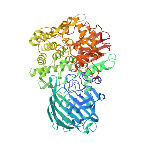
Lysosomal storage diseases are treated with human lysosomal enzymes produced in mammalian cells. Such enzyme therapeutics contain relatively low levels of mannose-6-phosphate, which is required to target them to the lysosomes of patient cells. Here we describe a method for increasing mannose-6-phosphate modification of lysosomal enzymes produced in yeast. We identified a glycosidase from C. cellulans that 'uncaps' N-glycans modified by yeast-type mannose-Pi-6-mannose to generate mammalian-type N-glycans with a mannose-6-phosphate substitution. Determination of the crystal structure of this glycosidase provided insight into its substrate specificity. We used this uncapping enzyme together with α-mannosidase to produce in yeast a form of the Pompe disease enzyme α-glucosidase rich in mannose-6-phosphate. Compared with the currently used therapeutic version, this form of α-glucosidase was more efficiently taken up by fibroblasts from Pompe disease patients, and it more effectively reduced cardiac muscular glycogen storage in a mouse model of the disease.
- Unit for Medical Biotechnology, Department for Molecular Biomedical Research, VIB, Ghent, Belgium.
Organizational Affiliation: