2XRY

X-ray structure of archaeal class II CPD photolyase from Methanosarcina mazei


Experimental Data Snapshot

  • Method: X-RAY DIFFRACTION
  • Resolution: 1.50 Å
  • R-Value Free: 0.210 
  • R-Value Work: 0.185 
  • R-Value Observed: 0.185 

wwPDB Validation   3D Report Full Report


Ligand Structure Quality Assessment 


This is version 1.2 of the entry. See complete history

Re-refinement Note

A newer entry is available that reflects an alternative modeling of the original data: 7F8T


Literature

Crystal Structures of an Archaeal Class II DNA Photolyase and its Complex with Uv-Damaged Duplex DNA.

Kiontke, S.Geisselbrecht, Y.Pokorny, R.Carell, T.Batschauer, A.Essen, L.O.

(2011) EMBO J 30: 4437

  • DOI: https://doi.org/10.1038/emboj.2011.313
  • Primary Citation of Related Structures:  
    2XRY, 2XRZ

  • PubMed Abstract: 

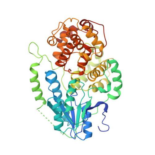
    Class II photolyases ubiquitously occur in plants, animals, prokaryotes and some viruses. Like the distantly related microbial class I photolyases, these enzymes repair UV-induced cyclobutane pyrimidine dimer (CPD) lesions within duplex DNA using blue/near-UV light. Methanosarcina mazei Mm0852 is a class II photolyase of the archaeal order of Methanosarcinales, and is closely related to plant and metazoan counterparts. Mm0852 catalyses light-driven DNA repair and photoreduction, but in contrast to class I enzymes lacks a high degree of binding discrimination between UV-damaged and intact duplex DNA. We solved crystal structures of Mm0852, the first one for a class II photolyase, alone and in complex with CPD lesion-containing duplex DNA. The lesion-binding mode differs from other photolyases by a larger DNA-binding site, and an unrepaired CPD lesion is found flipped into the active site and recognized by a cluster of five water molecules next to the bound 3'-thymine base. Different from other members of the photolyase-cryptochrome family, class II photolyases appear to utilize an unusual, conserved tryptophane dyad as electron transfer pathway to the catalytic FAD cofactor.


  • Organizational Affiliation

    Faculty of Chemistry, Department of Biochemistry, Philipps University, Marburg, Germany.


Macromolecules
Find similar proteins by:  (by identity cutoff)  |  3D Structure
Entity ID: 1
MoleculeChains Sequence LengthOrganismDetailsImage
DEOXYRIBODIPYRIMIDINE PHOTOLYASE482Methanosarcina mazei Go1Mutation(s): 1 
EC: 4.1.99.3
UniProt
Find proteins for Q8PYK9 (Methanosarcina mazei (strain ATCC BAA-159 / DSM 3647 / Goe1 / Go1 / JCM 11833 / OCM 88))
Explore Q8PYK9 
Go to UniProtKB:  Q8PYK9
Entity Groups  
Sequence Clusters30% Identity50% Identity70% Identity90% Identity95% Identity100% Identity
UniProt GroupQ8PYK9
Sequence Annotations
Expand
  • Reference Sequence
Small Molecules
Ligands 3 Unique
IDChains Name / Formula / InChI Key2D Diagram3D Interactions
FAD
Query on FAD

Download Ideal Coordinates CCD File 
B [auth A]FLAVIN-ADENINE DINUCLEOTIDE
C27 H33 N9 O15 P2
VWWQXMAJTJZDQX-UYBVJOGSSA-N
SO4
Query on SO4

Download Ideal Coordinates CCD File 
C [auth A]
D [auth A]
E [auth A]
F [auth A]
G [auth A]
C [auth A],
D [auth A],
E [auth A],
F [auth A],
G [auth A],
H [auth A],
I [auth A],
J [auth A],
K [auth A]
SULFATE ION
O4 S
QAOWNCQODCNURD-UHFFFAOYSA-L
GOL
Query on GOL

Download Ideal Coordinates CCD File 
L [auth A],
M [auth A]
GLYCEROL
C3 H8 O3
PEDCQBHIVMGVHV-UHFFFAOYSA-N
Experimental Data & Validation

Experimental Data

  • Method: X-RAY DIFFRACTION
  • Resolution: 1.50 Å
  • R-Value Free: 0.210 
  • R-Value Work: 0.185 
  • R-Value Observed: 0.185 
  • Space Group: P 43 21 2
Unit Cell:
Length ( Å )Angle ( ˚ )
a = 69.77α = 90
b = 69.77β = 90
c = 243.08γ = 90
Software Package:
Software NamePurpose
REFMACrefinement
XDSdata reduction
XSCALEdata scaling
PHASERphasing

Structure Validation

View Full Validation Report


Ligand Structure Quality Assessment 


Entry History 

Deposition Data

Revision History  (Full details and data files)

  • Version 1.0: 2011-09-14
    Type: Initial release
  • Version 1.1: 2011-11-16
    Changes: Database references
  • Version 1.2: 2024-05-01
    Changes: Data collection, Database references, Derived calculations, Other, Refinement description