2XR6

Crystal structure of the complex of the carbohydrate recognition domain of human DC-SIGN with pseudo trimannoside mimic.


Experimental Data Snapshot

  • Method: X-RAY DIFFRACTION
  • Resolution: 1.35 Å
  • R-Value Free: 0.168 
  • R-Value Work: 0.147 
  • R-Value Observed: 0.148 

wwPDB Validation   3D Report Full Report


Ligand Structure Quality Assessment 


This is version 1.6 of the entry. See complete history


Literature

Unique Dc-Sign Clustering Activity of a Small Glycomimetic: A Lesson for Ligand Design.

Sutkeviciute, I.Thepaut, M.Sattin, S.Berzi, A.Mcgeagh, J.Grudinin, S.Weiser, J.Le Roy, A.Reina, J.J.Rojo, J.Clerici, M.Bernardi, A.Ebel, C.Fieschi, F.

(2014) ACS Chem Biol 9: 1377

  • DOI: https://doi.org/10.1021/cb500054h
  • Primary Citation of Related Structures:  
    2XR6

  • PubMed Abstract: 

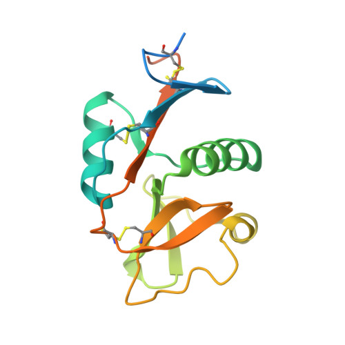
    DC-SIGN is a dendritic cell-specific C-type lectin receptor that recognizes highly glycosylated ligands expressed on the surface of various pathogens. This receptor plays an important role in the early stages of many viral infections, including HIV, which makes it an interesting therapeutic target. Glycomimetic compounds are good drug candidates for DC-SIGN inhibition due to their high solubility, resistance to glycosidases, and nontoxicity. We studied the structural properties of the interaction of the tetrameric DC-SIGN extracellular domain (ECD), with two glycomimetic antagonists, a pseudomannobioside (1) and a linear pseudomannotrioside (2). Though the inhibitory potency of 2, as measured by SPR competition experiments, was 1 order of magnitude higher than that of 1, crystal structures of the complexes within the DC-SIGN carbohydrate recognition domain showed the same binding mode for both compounds. Moreover, when conjugated to multivalent scaffolds, the inhibitory potencies of these compounds became uniform. Combining isothermal titration microcalorimetry, analytical ultracentrifugation, and dynamic light scattering techniques to study DC-SIGN ECD interaction with these glycomimetics revealed that 2 is able, without any multivalent presentation, to cluster DC-SIGN tetramers leading to an artificially overestimated inhibitory potency. The use of multivalent scaffolds presenting 1 or 2 in HIV trans-infection inhibition assay confirms the loss of potency of 2 upon conjugation and the equal efficacy of chemically simpler compound 1. This study documents a unique case where, among two active compounds chemically derived, the compound with the lower apparent activity is the optimal lead for further drug development.


  • Organizational Affiliation

    Univ. Grenoble Alpes, Institut de Biologie Structurale (IBS) , Grenoble F-38027, France.


Macromolecules
Find similar proteins by:  (by identity cutoff)  |  3D Structure
Entity ID: 1
MoleculeChains Sequence LengthOrganismDetailsImage
CD209 ANTIGEN170Homo sapiensMutation(s): 2 
UniProt & NIH Common Fund Data Resources
Find proteins for Q9NNX6 (Homo sapiens)
Explore Q9NNX6 
Go to UniProtKB:  Q9NNX6
PHAROS:  Q9NNX6
GTEx:  ENSG00000090659 
Entity Groups  
Sequence Clusters30% Identity50% Identity70% Identity90% Identity95% Identity100% Identity
UniProt GroupQ9NNX6
Sequence Annotations
Expand
  • Reference Sequence
Small Molecules
Ligands 5 Unique
IDChains Name / Formula / InChI Key2D Diagram3D Interactions
07B
Query on 07B

Download Ideal Coordinates CCD File 
C [auth A]dimethyl (1S,2S,4S,5S)-4,5-dihydroxycyclohexane-1,2-dicarboxylate
C10 H16 O6
ICVNZKRWDVXSLF-XAMCCFCMSA-N
MAN
Query on MAN

Download Ideal Coordinates CCD File 
B [auth A],
D [auth A]
alpha-D-mannopyranose
C6 H12 O6
WQZGKKKJIJFFOK-PQMKYFCFSA-N
AE9
Query on AE9

Download Ideal Coordinates CCD File 
E [auth A]2-AZIDOETHANOL
C2 H5 N3 O
BSULWPSUVMOMAN-UHFFFAOYSA-N
CA
Query on CA

Download Ideal Coordinates CCD File 
F [auth A],
G [auth A],
H [auth A]
CALCIUM ION
Ca
BHPQYMZQTOCNFJ-UHFFFAOYSA-N
CL
Query on CL

Download Ideal Coordinates CCD File 
I [auth A],
J [auth A],
K [auth A]
CHLORIDE ION
Cl
VEXZGXHMUGYJMC-UHFFFAOYSA-M
Experimental Data & Validation

Experimental Data

  • Method: X-RAY DIFFRACTION
  • Resolution: 1.35 Å
  • R-Value Free: 0.168 
  • R-Value Work: 0.147 
  • R-Value Observed: 0.148 
  • Space Group: P 43 21 2
Unit Cell:
Length ( Å )Angle ( ˚ )
a = 71.33α = 90
b = 71.33β = 90
c = 52.666γ = 90
Software Package:
Software NamePurpose
REFMACrefinement
XDSdata reduction
XDSdata scaling
MOLREPphasing

Structure Validation

View Full Validation Report


Ligand Structure Quality Assessment 


Entry History 

Deposition Data

Revision History  (Full details and data files)

  • Version 1.0: 2011-10-19
    Type: Initial release
  • Version 1.1: 2014-07-30
    Changes: Database references, Non-polymer description
  • Version 1.2: 2014-08-27
    Changes: Database references
  • Version 1.3: 2019-01-30
    Changes: Data collection, Experimental preparation, Other
  • Version 1.4: 2019-03-06
    Changes: Data collection, Derived calculations, Experimental preparation
  • Version 1.5: 2020-07-29
    Type: Remediation
    Reason: Carbohydrate remediation
    Changes: Data collection, Derived calculations, Other, Structure summary
  • Version 1.6: 2023-12-20
    Changes: Data collection, Database references, Derived calculations, Refinement description, Structure summary