2XNK

Structure and function of the Rad9-binding region of the DNA damage checkpoint adaptor TopBP1


Experimental Data Snapshot

  • Method: X-RAY DIFFRACTION
  • Resolution: 2.60 Å
  • R-Value Free: 0.266 
  • R-Value Work: 0.205 
  • R-Value Observed: 0.208 

wwPDB Validation   3D Report Full Report


This is version 1.3 of the entry. See complete history


Literature

Structure and Function of the Rad9-Binding Region of the DNA-Damage Checkpoint Adaptor Topbp1.

Rappas, M.Oliver, A.W.Pearl, L.H.

(2011) Nucleic Acids Res 39: 313

  • DOI: https://doi.org/10.1093/nar/gkq743
  • Primary Citation of Related Structures:  
    2XNH, 2XNK

  • PubMed Abstract: 

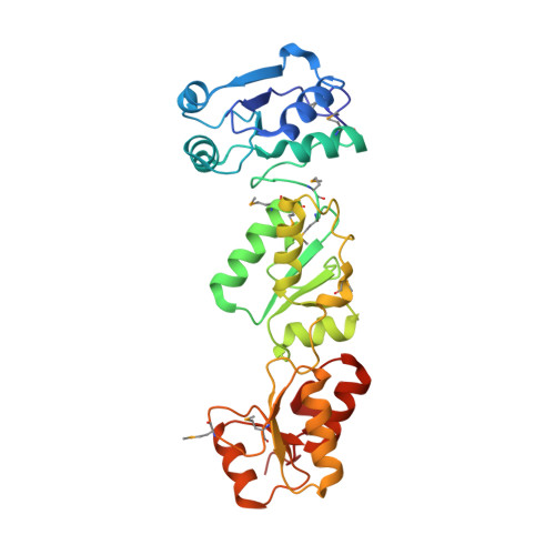
    TopBP1 is a scaffold protein that coordinates activation of the DNA-damage-checkpoint response by coupling binding of the 9-1-1 checkpoint clamp at sites of ssDNA, to activation of the ATR-ATRIP checkpoint kinase complex. We have now determined the crystal structure of the N-terminal region of human TopBP1, revealing an unexpected triple-BRCT domain structure. The arrangement of the BRCT domains differs significantly from previously described tandem BRCT domain structures, and presents two distinct sites for binding phosphopeptides in the second and third BRCT domains. We show that the site in the second but not third BRCT domain in the N-terminus of TopBP1, provides specific interaction with a phosphorylated motif at pSer387 in Rad9, which can be generated by CK2.


  • Organizational Affiliation

    Cancer Research UK DNA Repair Enzyme Group, Section of Structural Biology, The Institute of Cancer Research, London, UK.


Macromolecules
Find similar proteins by:  (by identity cutoff)  |  3D Structure
Entity ID: 1
MoleculeChains Sequence LengthOrganismDetailsImage
DNA TOPOISOMERASE 2-BINDING PROTEIN 1
A, B, C, D
292Homo sapiensMutation(s): 0 
UniProt & NIH Common Fund Data Resources
Find proteins for Q92547 (Homo sapiens)
Explore Q92547 
Go to UniProtKB:  Q92547
PHAROS:  Q92547
GTEx:  ENSG00000163781 
Entity Groups  
Sequence Clusters30% Identity50% Identity70% Identity90% Identity95% Identity100% Identity
UniProt GroupQ92547
Sequence Annotations
Expand
  • Reference Sequence
Small Molecules
Ligands 1 Unique
IDChains Name / Formula / InChI Key2D Diagram3D Interactions
GOL
Query on GOL

Download Ideal Coordinates CCD File 
E [auth A]
F [auth A]
G [auth B]
H [auth B]
I [auth B]
E [auth A],
F [auth A],
G [auth B],
H [auth B],
I [auth B],
J [auth C],
K [auth C],
L [auth C],
M [auth D],
N [auth D],
O [auth D]
GLYCEROL
C3 H8 O3
PEDCQBHIVMGVHV-UHFFFAOYSA-N
Modified Residues  1 Unique
IDChains TypeFormula2D DiagramParent
MSE
Query on MSE
A, B, C, D
L-PEPTIDE LINKINGC5 H11 N O2 SeMET
Experimental Data & Validation

Experimental Data

  • Method: X-RAY DIFFRACTION
  • Resolution: 2.60 Å
  • R-Value Free: 0.266 
  • R-Value Work: 0.205 
  • R-Value Observed: 0.208 
  • Space Group: P 1 21 1
Unit Cell:
Length ( Å )Angle ( ˚ )
a = 39.49α = 90
b = 290.28β = 89.85
c = 60.58γ = 90
Software Package:
Software NamePurpose
PHENIXrefinement
MOSFLMdata reduction
SCALAdata scaling
PHASERphasing

Structure Validation

View Full Validation Report


Entry History 

Deposition Data

Revision History  (Full details and data files)

  • Version 1.0: 2010-09-01
    Type: Initial release
  • Version 1.1: 2011-05-08
    Changes: Version format compliance
  • Version 1.2: 2011-07-13
    Changes: Version format compliance
  • Version 1.3: 2023-12-20
    Changes: Data collection, Database references, Derived calculations, Other, Refinement description