2XN0 | pdb_00002xn0

Structure of alpha-galactosidase from Lactobacillus acidophilus NCFM, PtCl4 derivative


Experimental Data Snapshot

  • Method: X-RAY DIFFRACTION
  • Resolution: 2.50 Å
  • R-Value Free: 
    0.203 (Depositor), 0.200 (DCC) 
  • R-Value Work: 
    0.148 (Depositor), 0.146 (DCC) 
  • R-Value Observed: 
    0.150 (Depositor) 

wwPDB Validation 3D Report Full Report

Validation slider image for 2XN0

This is version 1.4 of the entry. See complete history

Literature

Crystal Structure of Alpha-Galactosidase from Lactobacillus Acidophilus Ncfm: Insight Into Tetramer Formation and Substrate Binding.

Fredslund, F.Abou Hachem, M.Jonsgaard Larsen, R.Gerd Sorensen, P.Coutinho, P.M.Lo Leggio, L.Svensson, B.

(2011) J Mol Biology 412: 466

  • DOI: https://doi.org/10.1016/j.jmb.2011.07.057
  • Primary Citation Related Structures: 
    2XN0, 2XN1, 2XN2

  • PubMed Abstract: 

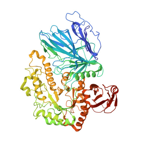
    Lactobacillus acidophilus NCFM is a probiotic bacterium known for its beneficial effects on human health. The importance of α-galactosidases (α-Gals) for growth of probiotic organisms on oligosaccharides of the raffinose family present in many foods is increasingly recognized. Here, the crystal structure of α-Gal from L. acidophilus NCFM (LaMel36A) of glycoside hydrolase (GH) family 36 (GH36) is determined by single-wavelength anomalous dispersion. In addition, a 1.58-Å-resolution crystallographic complex with α-d-galactose at substrate binding subsite -1 was determined. LaMel36A has a large N-terminal twisted β-sandwich domain, connected by a long α-helix to the catalytic (β/α)(8)-barrel domain, and a C-terminal β-sheet domain. Four identical monomers form a tightly packed tetramer where three monomers contribute to the structural integrity of the active site in each monomer. Structural comparison of LaMel36A with the monomeric Thermotoga maritima α-Gal (TmGal36A) reveals that O2 of α-d-galactose in LaMel36A interacts with a backbone nitrogen in a glycine-rich loop of the catalytic domain, whereas the corresponding atom in TmGal36A is from a tryptophan side chain belonging to the N-terminal domain. Thus, two distinctly different structural motifs participate in substrate recognition. The tetrameric LaMel36A furthermore has a much deeper active site than the monomeric TmGal36A, which possibly modulates substrate specificity. Sequence analysis of GH36, inspired by the observed structural differences, results in four distinct subgroups having clearly different active-site sequence motifs. This novel subdivision incorporates functional and architectural features and may aid further biochemical and structural analyses within GH36.


  • Organizational Affiliation
    • Department of Systems Biology, Enzyme and Protein Chemistry, Technical University of Denmark, Søltofts Plads, Building 224, DK-2800 Kongens Lyngby, Denmark.

Macromolecule Content 

  • Total Structure Weight: 173.69 kDa 
  • Atom Count: 12,849 
  • Modeled Residue Count: 1,458 
  • Deposited Residue Count: 1,464 
  • Unique protein chains: 1

Macromolecules

Find similar proteins by:|  3D Structure
Entity ID: 1
MoleculeChains  Sequence LengthOrganismDetailsImage
ALPHA-GALACTOSIDASE
A, B
732Lactobacillus acidophilus NCFMMutation(s): 0 
EC: 3.2.1.22
UniProt
Find proteins for G1UB44 (Lactobacillus acidophilus (strain ATCC 700396 / NCK56 / N2 / NCFM))
Explore G1UB44 
Go to UniProtKB:  G1UB44
Entity Groups
Sequence Clusters30% Identity50% Identity70% Identity90% Identity95% Identity100% Identity
UniProt GroupG1UB44
Sequence Annotations
Expand
Reference Sequence

Small Molecules

Ligands 2 Unique
IDChains Name / Formula / InChI Key2D Diagram3D Interactions
PT

Query on PT


Download:Ideal Coordinates CCD File
C [auth A]
D [auth A]
E [auth A]
F [auth A]
G [auth A]
C [auth A],
D [auth A],
E [auth A],
F [auth A],
G [auth A],
H [auth A],
I [auth A],
J [auth A],
R [auth A],
S [auth B],
T [auth B],
U [auth B],
V [auth B],
W [auth B],
X [auth B],
Y [auth B],
Z [auth B]
PLATINUM (II) ION
Pt
HRGDZIGMBDGFTC-UHFFFAOYSA-N
GOL

Query on GOL


Download:Ideal Coordinates CCD File
AA [auth B]
BA [auth B]
CA [auth B]
DA [auth B]
EA [auth B]
AA [auth B],
BA [auth B],
CA [auth B],
DA [auth B],
EA [auth B],
FA [auth B],
K [auth A],
L [auth A],
M [auth A],
N [auth A],
O [auth A],
P [auth A],
Q [auth A]
GLYCEROL
C3 H8 O3
PEDCQBHIVMGVHV-UHFFFAOYSA-N

Experimental Data & Validation

Experimental Data

  • Method: X-RAY DIFFRACTION
  • Resolution: 2.50 Å
  • R-Value Free:  0.203 (Depositor), 0.200 (DCC) 
  • R-Value Work:  0.148 (Depositor), 0.146 (DCC) 
  • R-Value Observed: 0.150 (Depositor) 
Space Group: P 21 21 2
Unit Cell:
Length ( Å )Angle ( ˚ )
a = 126.37α = 90
b = 149.58β = 90
c = 90.84γ = 90
Software Package:
Software NamePurpose
PHENIXrefinement
XDSdata reduction
XSCALEdata scaling
PHENIXphasing

Structure Validation

View Full Validation Report


Entry History 

Deposition Data

Revision History  (Full details and data files)

  • Version 1.0: 2011-08-10
    Type: Initial release
  • Version 1.1: 2011-08-31
    Changes: Database references, Structure summary
  • Version 1.2: 2011-09-14
    Changes: Database references, Refinement description
  • Version 1.3: 2017-07-12
    Changes: Data collection, Refinement description
  • Version 1.4: 2024-05-08
    Changes: Data collection, Database references, Derived calculations, Other