Distal-to-Proximal No Conversion in Hemoproteins: The Role of the Proximal Pocket.
Hough, M.A., Antonyuk, S.V., Barbieri, S., Rustage, N., Mckay, A.L., Servid, A.E., Eady, R.R., Andrew, C.R., Hasnain, S.S.(2011) J Mol Biology 405: 395
- PubMed: 21073879 Search on PubMed
- DOI: https://doi.org/10.1016/j.jmb.2010.10.035
- Primary Citation Related Structures:
2XL6, 2XL8, 2XLD, 2XLE, 2XLH, 2XLM, 2XLO, 2XLV, 2XLW, 2XM0, 2XM4 - PubMed Abstract:
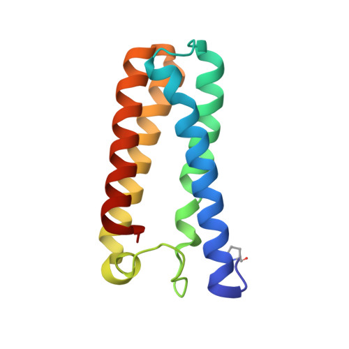
Hemoproteins play central roles in the formation and utilization of nitric oxide (NO) in cellular signaling, as well as in protection against nitrosative stress. Key to heme-nitrosyl function and reactivity is the Fe coordination number (5 or 6). For (five-coordinate) 5c-NO complexes, the potential for NO to bind on either heme face exists, as in the microbial cytochrome c' from Alcaligenes xylosoxidans (AxCYTcp), which forms a stable proximal 5c-NO complex via a distal six-coordinate NO intermediate and a putative dinitrosyl species. Strong parallels between the NO-binding kinetics of AxCYTcp, the eukaryotic NO sensor soluble guanylate cyclase, and the ferrocytochrome c/cardiolipin complex have led to the suggestion that a distal-to-proximal NO switch could contribute to the selective ligand responses in gas-sensing hemoproteins. The proximal NO-binding site in AxCYTcp is close to a conserved basic (Arg124) residue that is postulated to modulate NO reactivity. We have replaced Arg124 by five different amino acids and have determined high-resolution (1.07-1.40 Å) crystallographic structures with and without NO. These, together with kinetic and resonance Raman data, provide new insights into the mechanism of distal-to-proximal heme-NO conversion, including the determinants of Fe-His bond scission. The Arg124Ala variant allowed us to determine the structure of an analog of the previously unobserved key 5c-NO distal intermediate species. The very high resolution structures combined with the extensive spectroscopic and kinetic data have allowed us to provide a fresh insight into heme reactivity towards NO, a reaction that is of wide importance in biology.
- Faculty of Health and Life Sciences, University of Liverpool, Liverpool L69 7ZB, UK.
Organizational Affiliation: