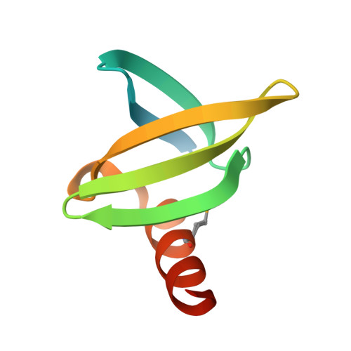
Tolerance of the Archaeal Sac7D Scaffold Protein to Alternative Library Designs: Characterization of Anti-Immunoglobulin G Affitins.
Behar, G., Bellinzoni, M., Maillasson, M., Paillard-Laurance, L., Alzari, P.M., He, X., Mouratou, B., Pecorari, F.(2013) Protein Eng Des Sel 26: 267
- PubMed: 23315487 Search on PubMed
- DOI: https://doi.org/10.1093/protein/gzs106
- Primary Citation Related Structures:
2XIW - PubMed Abstract:
Engineered protein scaffolds have received considerable attention as alternatives to antibodies in both basic and applied research, as they can offer superior biophysical properties often associated with a simpler molecular organization. Sac7d has been demonstrated as an effective scaffold for molecular recognition. Here, we used the initial L1 'flat surface' library constructed by randomization of 14 residues, to identify ligands specific for human immunoglobulin G. To challenge the plasticity of the Sac7d protein scaffold, we designed the alternative L2 'flat surface & loops' library whereof only 10 residues are randomized. Representative binders (Affitins) of the two libraries exhibited affinities in the low nanomolar range and were able to recognize different epitopes within human immunoglobulin G. These Affitins were stable up to pH 12 while largely conserving other favorable properties of Sac7d protein, such as high expression yields in Escherichia coli, solubility, thermal stability up to 80.7°C, and acidic stability (pH 0). In agreement with our library designs, mutagenesis study revealed two distinct binding areas, one including loops. Together, our results indicate that the Sac7d scaffold tolerates alternative library designs, which further expands the diversity of Affitins and may provide a general way to create tailored affinity tools for demanding applications.
- Université de Nantes, UMR CNRS 6204, Ingénierie de la reconnaissance, F-44322 Nantes, France.
Organizational Affiliation: