Structural and Functional Characterization of Sporosag: A Sag2 Related Surface Antigen from Toxoplasma Gondii.
Crawford, J., Lamb, E., Wasmuth, J., Grujic, O., Grigg, M.E., Boulanger, M.J.(2010) J Biological Chem 285: 12063
- PubMed: 20164173 Search on PubMedSearch on PubMed Central
- DOI: https://doi.org/10.1074/jbc.M109.054866
- Primary Citation Related Structures:
2WNK, 2X28 - PubMed Abstract:
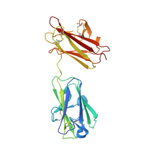
Toxoplasma gondii, the etiological agent of toxoplasmosis, utilizes stage-specific expression of antigenically distinct glycosylphosphatidylinositol-tethered surface coat proteins to promote and establish chronic infection. Of the three infective stages of T. gondii, sporozoites are encapsulated in highly infectious oocysts that have been linked to large scale outbreaks of toxoplasmosis. SporoSAG (surface antigen glycoprotein) is the dominant surface coat protein expressed on the surface of sporozoites. Using a bioinformatic approach, we show that SporoSAG clusters with the SAG2 subfamily of the SAG1-related superfamily (SRS) and is non-polymorphic among the 11 haplogroups of T. gondii strains. In contrast to the immunodominant SAG1 protein expressed on tachyzoites, SporoSAG is non-immunogenic during natural infection. We report the 1.60 A resolution crystal structure of SporoSAG solved using cadmium single anomalous dispersion. SporoSAG crystallized as a monomer and displays unique features of the SRS beta-sandwich fold relative to SAG1 and BSR4. Intriguingly, the structural diversity is localized to the upper sheets of the beta-sandwich fold and may have important implications for multimerization and host cell ligand recognition. The structure of SporoSAG also reveals an unexpectedly acidic surface that contrasts with the previously determined SAG1 and BSR4 structures where a basic surface is predicted to play a role in binding negatively charged glycosaminoglycans. Our structural and functional characterization of SporoSAG provides a rationale for the evolutionary divergence of this key SRS family member.
- Department of Biochemistry and Microbiology, University of Victoria, Victoria, British Columbia V8W 3P6, Canada.
Organizational Affiliation: