2WYC | pdb_00002wyc

The quorum quenching N-acyl homoserine lactone acylase PvdQ in complex with 3-oxo-lauric acid


Experimental Data Snapshot

  • Method: X-RAY DIFFRACTION
  • Resolution: 1.90 Å
  • R-Value Free: 
    0.190 (Depositor), 0.209 (DCC) 
  • R-Value Work: 
    0.163 (Depositor), 0.186 (DCC) 
  • R-Value Observed: 
    0.164 (Depositor) 

wwPDB Validation 3D Report Full Report

Validation slider image for 2WYC

Ligand Structure Quality Assessment 


This is version 1.3 of the entry. See complete history

Literature

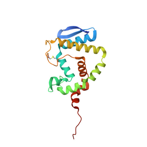
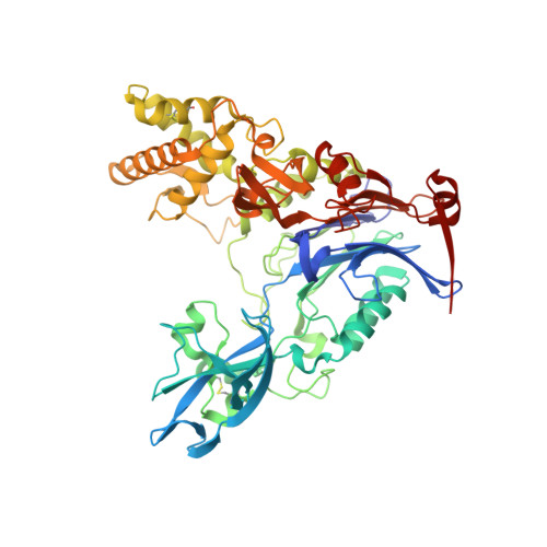
The Quorum-Quenching N-Acyl Homoserine Lactone Acylase Pvdq is an Ntn-Hydrolase with an Unusual Substrate-Binding Pocket

Bokhove, M.Nadal Jimenez, P.Quax, W.J.Dijkstra, B.W.

(2010) Proc Natl Acad Sci U S A 107: 686

  • DOI: https://doi.org/10.1073/pnas.0911839107
  • Primary Citation Related Structures: 
    2WYB, 2WYC, 2WYD, 2WYE

  • PubMed Abstract: 

    In many Gram-negative pathogens, their virulent behavior is regulated by quorum sensing, in which diffusible signals such as N-acyl homoserine lactones (AHLs) act as chemical messaging compounds. Enzymatic degradation of these diffusible signals by, e.g., lactonases or amidohydrolases abolishes AHL regulated virulence, a process known as quorum quenching. Here we report the first crystal structure of an AHL amidohydrolase, the AHL acylase PvdQ from Pseudomonas aeruginosa. PvdQ has a typical alpha/beta heterodimeric Ntn-hydrolase fold, similar to penicillin G acylase and cephalosporin acylase. However, it has a distinct, unusually large, hydrophobic binding pocket, ideally suited to recognize C12 fatty acid-like chains of AHLs. Binding of a C12 fatty acid or a 3-oxo-C12 fatty acid induces subtle conformational changes to accommodate the aliphatic chain. Furthermore, the structure of a covalent ester intermediate identifies Serbeta1 as the nucleophile and Asnbeta269 and Valbeta70 as the oxyanion hole residues in the AHL degradation process. Our structures show the versatility of the Ntn-hydrolase scaffold and can serve as a structural paradigm for Ntn-hydrolases with similar substrate preference. Finally, the quorum-quenching capabilities of PvdQ may be utilized to suppress the quorum-sensing machinery of pathogens.


  • Organizational Affiliation
    • Laboratory of Biophysical Chemistry, University of Groningen, Nijenborgh 4, 9747 AG Groningen, The Netherlands.

Macromolecule Content 

  • Total Structure Weight: 80.2 kDa 
  • Atom Count: 6,094 
  • Modeled Residue Count: 710 
  • Deposited Residue Count: 716 
  • Unique protein chains: 2

Macromolecules

Find similar proteins by:|  3D Structure
Entity ID: 1
MoleculeChains  Sequence LengthOrganismDetailsImage
ACYL-HOMOSERINE LACTONE ACYLASE PVDQ SUBUNIT ALPHA170Pseudomonas aeruginosa PAO1Mutation(s): 0 
EC: 3.5.1.97
UniProt
Find proteins for Q9I194 (Pseudomonas aeruginosa (strain ATCC 15692 / DSM 22644 / CIP 104116 / JCM 14847 / LMG 12228 / 1C / PRS 101 / PAO1))
Explore Q9I194 
Go to UniProtKB:  Q9I194
Entity Groups
Sequence Clusters30% Identity50% Identity70% Identity90% Identity95% Identity100% Identity
UniProt GroupQ9I194
Sequence Annotations
Expand
Reference Sequence
Find similar proteins by:|  3D Structure
Entity ID: 2
MoleculeChains  Sequence LengthOrganismDetailsImage
ACYL-HOMOSERINE LACTONE ACYLASE PVDQ SUBUNIT BETA546Pseudomonas aeruginosa PAO1Mutation(s): 0 
EC: 3.5.1.97
UniProt
Find proteins for Q9I194 (Pseudomonas aeruginosa (strain ATCC 15692 / DSM 22644 / CIP 104116 / JCM 14847 / LMG 12228 / 1C / PRS 101 / PAO1))
Explore Q9I194 
Go to UniProtKB:  Q9I194
Entity Groups
Sequence Clusters30% Identity50% Identity70% Identity90% Identity95% Identity100% Identity
UniProt GroupQ9I194
Sequence Annotations
Expand
Reference Sequence

Small Molecules

Ligands 3 Unique
IDChains Name / Formula / InChI Key2D Diagram3D Interactions
3LA

Query on 3LA


Download:Ideal Coordinates CCD File
M [auth B]3-OXODODECANOIC ACID
C12 H22 O3
DZHSPYMHDVROSM-UHFFFAOYSA-N
GOL

Query on GOL


Download:Ideal Coordinates CCD File
C [auth A]
D [auth A]
E [auth A]
F [auth A]
G [auth A]
C [auth A],
D [auth A],
E [auth A],
F [auth A],
G [auth A],
H [auth A],
I [auth B],
J [auth B],
K [auth B]
GLYCEROL
C3 H8 O3
PEDCQBHIVMGVHV-UHFFFAOYSA-N
DMS

Query on DMS


Download:Ideal Coordinates CCD File
L [auth B]DIMETHYL SULFOXIDE
C2 H6 O S
IAZDPXIOMUYVGZ-UHFFFAOYSA-N

Experimental Data & Validation

Experimental Data

  • Method: X-RAY DIFFRACTION
  • Resolution: 1.90 Å
  • R-Value Free:  0.190 (Depositor), 0.209 (DCC) 
  • R-Value Work:  0.163 (Depositor), 0.186 (DCC) 
  • R-Value Observed: 0.164 (Depositor) 
Space Group: C 2 2 21
Unit Cell:
Length ( Å )Angle ( ˚ )
a = 120.8α = 90
b = 166.5β = 90
c = 94.3γ = 90
Software Package:
Software NamePurpose
REFMACrefinement
XDSdata reduction
SCALAdata scaling
PHASERphasing

Structure Validation

View Full Validation Report


Ligand Structure Quality Assessment 


Entry History 

Deposition Data

Revision History  (Full details and data files)

  • Version 1.0: 2009-12-29
    Type: Initial release
  • Version 1.1: 2011-05-08
    Changes: Version format compliance
  • Version 1.2: 2011-07-13
    Changes: Version format compliance
  • Version 1.3: 2024-11-13
    Changes: Data collection, Database references, Derived calculations, Other, Structure summary