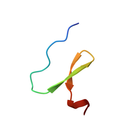
2WW6

foldon containing D-amino acids in turn positions


Experimental Data Snapshot

  • Method: X-RAY DIFFRACTION
  • Resolution: 0.98 Å
  • R-Value Free: 0.147 
  • R-Value Work: 0.129 
  • R-Value Observed: 0.130 

Starting Model: experimental
View more details

wwPDB Validation   3D Report Full Report


This is version 1.4 of the entry. See complete history


Literature

Structural Characterization of a Beta-Turn Mimic within a Protein-Protein Interface.

Eckhardt, B.Grosse, W.Essen, L.Geyer, A.

(2010) Proc Natl Acad Sci U S A 107: 18336

  • DOI: https://doi.org/10.1073/pnas.1004187107
  • Primary Citation of Related Structures:  
    2WW6, 2WW7

  • PubMed Abstract: 

    β-Turns are secondary structure elements not only exposed on protein surfaces, but also frequently found to be buried in protein-protein interfaces. Protein engineering so far considered mainly the backbone-constraining properties of synthetic β-turn mimics as parts of surface-exposed loops. A β-turn mimic, Hot═Tap, that is available in gram amounts, provides two hydroxyl groups that enhance its turn-inducing properties besides being able to form side-chain-like interactions. NMR studies on cyclic hexapeptides harboring the Hot═Tap dipeptide proved its strong β-turn-inducing capability. Crystallographic analyses of the trimeric fibritin-foldon/Hot═Tap hybrid reveal at atomic resolution how Hot═Tap replaces a βI'-turn by a βII'-type structure. Furthermore, Hot═Tap adapts to the complex protein environment by participating in several direct and water-bridged interactions across the foldon trimer interface. As building blocks, β-turn mimics capable of both backbone and side-chain mimicry may simplify the design of synthetic proteins.


  • Organizational Affiliation

    Fachbereich Chemie, Philipps-Universität Marburg, Hans-Meerwein-Strasse, D-35032 Marburg, Germany.


Macromolecules
Find similar proteins by:  (by identity cutoff)  |  3D Structure
Entity ID: 1
MoleculeChains Sequence LengthOrganismDetailsImage
FIBRITIN
A, B, C
27Tequatrovirus T4Mutation(s): 0 
UniProt
Find proteins for Q76VI8 (Tequatrovirus T2)
Explore Q76VI8 
Go to UniProtKB:  Q76VI8
Entity Groups  
Sequence Clusters30% Identity50% Identity70% Identity90% Identity95% Identity100% Identity
UniProt GroupQ76VI8
Sequence Annotations
Expand
  • Reference Sequence
Small Molecules
Ligands 1 Unique
IDChains Name / Formula / InChI Key2D Diagram3D Interactions
PG4
Query on PG4

Download Ideal Coordinates CCD File 
D [auth C]TETRAETHYLENE GLYCOL
C8 H18 O5
UWHCKJMYHZGTIT-UHFFFAOYSA-N
Experimental Data & Validation

Experimental Data

  • Method: X-RAY DIFFRACTION
  • Resolution: 0.98 Å
  • R-Value Free: 0.147 
  • R-Value Work: 0.129 
  • R-Value Observed: 0.130 
  • Space Group: P 32
Unit Cell:
Length ( Å )Angle ( ˚ )
a = 29.83α = 90
b = 29.83β = 90
c = 75.08γ = 120
Software Package:
Software NamePurpose
REFMACrefinement
MOSFLMdata reduction
SCALAdata scaling
PHASERphasing

Structure Validation

View Full Validation Report


Entry History 

Deposition Data

Revision History  (Full details and data files)

  • Version 1.0: 2010-09-29
    Type: Initial release
  • Version 1.1: 2011-05-08
    Changes: Version format compliance
  • Version 1.2: 2011-07-13
    Changes: Version format compliance
  • Version 1.3: 2023-12-20
    Changes: Data collection, Database references, Derived calculations, Other, Refinement description
  • Version 1.4: 2024-11-06
    Changes: Structure summary