2WSS

The structure of the membrane extrinsic region of bovine ATP synthase


Experimental Data Snapshot

  • Method: X-RAY DIFFRACTION
  • Resolution: 3.20 Å
  • R-Value Free: 0.271 
  • R-Value Work: 0.217 
  • R-Value Observed: 0.220 

wwPDB Validation   3D Report Full Report


Ligand Structure Quality Assessment 


This is version 1.3 of the entry. See complete history


Literature

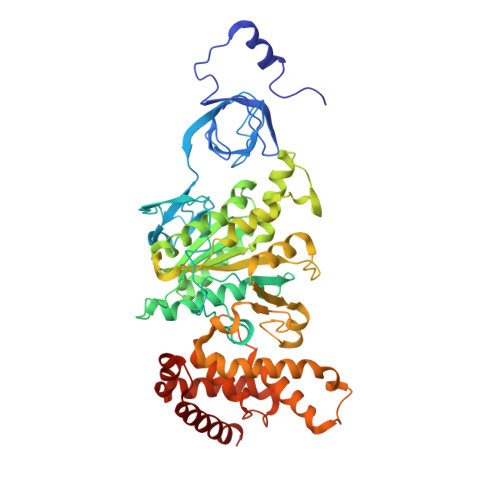
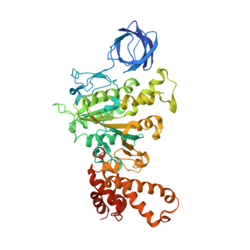
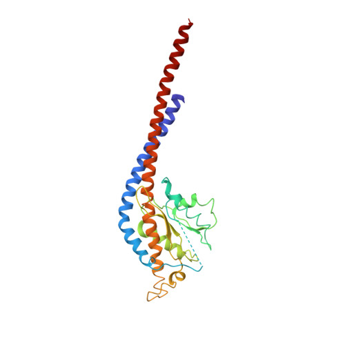
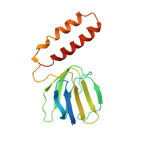
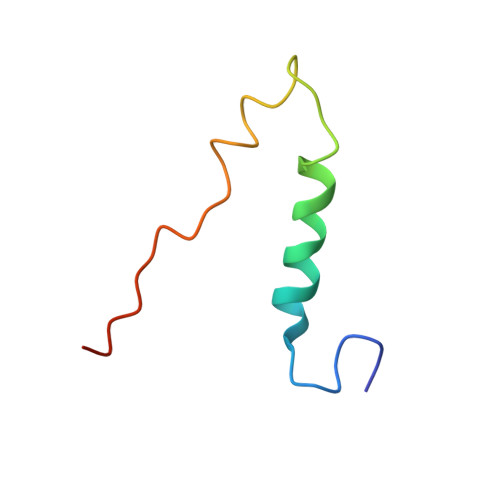
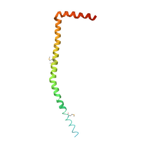
The Structure of the Membrane Extrinsic Region of Bovine ATP Synthase

Rees, D.M.Leslie, A.G.W.Walker, J.E.

(2009) Proc Natl Acad Sci U S A 106: 21597

  • DOI: https://doi.org/10.1073/pnas.0910365106
  • Primary Citation of Related Structures:  
    2WSS

  • PubMed Abstract: 
  • The structure of the complex between bovine mitochondrial F(1)-ATPase and a stator subcomplex has been determined at a resolution of 3.2 A. The resolved region of the stator contains residues 122-207 of subunit b; residues 5-25 and 35-57 of F(6); 3 segments of subunit d from residues 30-40, 65-74, and 85-91; and residues 1-146 and 169-189 of the oligomycin sensitivity conferral protein (OSCP) ...

    The structure of the complex between bovine mitochondrial F(1)-ATPase and a stator subcomplex has been determined at a resolution of 3.2 A. The resolved region of the stator contains residues 122-207 of subunit b; residues 5-25 and 35-57 of F(6); 3 segments of subunit d from residues 30-40, 65-74, and 85-91; and residues 1-146 and 169-189 of the oligomycin sensitivity conferral protein (OSCP). The stator subcomplex represents its membrane distal part, and its structure has been augmented with an earlier structure of a subcomplex containing residues 79-183, 3-123, and 5-70 of subunits b, d, and F(6), respectively, which extends to the surface of the inner membrane of the mitochondrion. The N-terminal domain of the OSCP links the stator with F(1)-ATPase via alpha-helical interactions with the N-terminal region of subunit alpha(E). Its C-terminal domain makes extensive helix-helix interactions with the C-terminal alpha-helix of subunit b from residues 190-207. Subunit b extends as a continuous 160-A long alpha-helix from residue 188 back to residue 79 near to the surface of the inner mitochondrial membrane. This helix appears to be stiffened by other alpha-helices in subunits d and F(6), but the structure can bend inward toward the F(1) domain around residue 146 of subunit b. The linker region between the 2 domains of the OSCP also appears to be flexible, enabling the stator to adjust its shape as it passes over the changing profile of the F(1) domain during a catalytic cycle. The structure of the membrane extrinsic part of bovine ATP synthase is now complete.


    Organizational Affiliation

    Medical Research Council Mitochondrial Biology Unit, Wellcome Trust/Medical Research Council Building, Hills Road, Cambridge CB2 0XY, United Kingdom.


Macromolecules
Find similar proteins by:  (by identity cutoff)  |  3D Structure
Entity ID: 1
MoleculeChainsSequence LengthOrganismDetailsImage
ATP SYNTHASE SUBUNIT ALPHA, MITOCHONDRIAL
A, B, C, J, K
A, B, C, J, K, L
510Bos taurusMutation(s): 0 
Gene Names: ATP5F1AATP5A1ATP5A2
Membrane Entity: Yes 
UniProt
Find proteins for P19483 (Bos taurus)
Explore P19483 
Go to UniProtKB:  P19483
Entity Groups  
Sequence Clusters30% Identity50% Identity70% Identity90% Identity95% Identity100% Identity
UniProt GroupP19483
Protein Feature View
Expand
  • Reference Sequence
Find similar proteins by:  (by identity cutoff)  |  3D Structure
Entity ID: 2
MoleculeChainsSequence LengthOrganismDetailsImage
ATP SYNTHASE SUBUNIT BETA, MITOCHONDRIAL
D, E, F, M, N
D, E, F, M, N, O
482Bos taurusMutation(s): 0 
Gene Names: ATP5F1BATP5B
EC: 3.6.3.14 (PDB Primary Data), 7.1.2.2 (UniProt)
Membrane Entity: Yes 
UniProt
Find proteins for P00829 (Bos taurus)
Explore P00829 
Go to UniProtKB:  P00829
Entity Groups  
Sequence Clusters30% Identity50% Identity70% Identity90% Identity95% Identity100% Identity
UniProt GroupP00829
Protein Feature View
Expand
  • Reference Sequence
Find similar proteins by:  (by identity cutoff)  |  3D Structure
Entity ID: 3
MoleculeChainsSequence LengthOrganismDetailsImage
ATP SYNTHASE SUBUNIT GAMMA, MITOCHONDRIAL
G, P
272Bos taurusMutation(s): 0 
Gene Names: ATP5F1CATP5CATP5C1
Membrane Entity: Yes 
UniProt
Find proteins for P05631 (Bos taurus)
Explore P05631 
Go to UniProtKB:  P05631
Entity Groups  
Sequence Clusters30% Identity50% Identity70% Identity90% Identity95% Identity100% Identity
UniProt GroupP05631
Protein Feature View
Expand
  • Reference Sequence
Find similar proteins by:  (by identity cutoff)  |  3D Structure
Entity ID: 4
MoleculeChainsSequence LengthOrganismDetailsImage
ATP SYNTHASE SUBUNIT DELTA, MITOCHONDRIAL
H, Q
146Bos taurusMutation(s): 0 
Gene Names: ATP5F1DATP5D
Membrane Entity: Yes 
UniProt
Find proteins for P05630 (Bos taurus)
Explore P05630 
Go to UniProtKB:  P05630
Entity Groups  
Sequence Clusters30% Identity50% Identity70% Identity90% Identity95% Identity100% Identity
UniProt GroupP05630
Protein Feature View
Expand
  • Reference Sequence
Find similar proteins by:  (by identity cutoff)  |  3D Structure
Entity ID: 5
MoleculeChainsSequence LengthOrganismDetailsImage
ATP SYNTHASE SUBUNIT EPSILON, MITOCHONDRIAL
I, R
50Bos taurusMutation(s): 0 
Gene Names: ATP5F1EATP5E
Membrane Entity: Yes 
UniProt
Find proteins for P05632 (Bos taurus)
Explore P05632 
Go to UniProtKB:  P05632
Entity Groups  
Sequence Clusters30% Identity50% Identity70% Identity90% Identity95% Identity100% Identity
UniProt GroupP05632
Protein Feature View
Expand
  • Reference Sequence
Find similar proteins by:  (by identity cutoff)  |  3D Structure
Entity ID: 6
MoleculeChainsSequence LengthOrganismDetailsImage
ATP SYNTHASE SUBUNIT O, MITOCHONDRIAL
S, W
190Bos taurusMutation(s): 0 
Gene Names: ATP5POATP5O
Membrane Entity: Yes 
UniProt
Find proteins for P13621 (Bos taurus)
Explore P13621 
Go to UniProtKB:  P13621
Entity Groups  
Sequence Clusters30% Identity50% Identity70% Identity90% Identity95% Identity100% Identity
UniProt GroupP13621
Protein Feature View
Expand
  • Reference Sequence
Find similar proteins by:  (by identity cutoff)  |  3D Structure
Entity ID: 7
MoleculeChainsSequence LengthOrganismDetailsImage
ATP SYNTHASE SUBUNIT B, MITOCHONDRIAL
T, X
116Bos taurusMutation(s): 0 
Gene Names: ATP5PBATP5FATP5F1
Membrane Entity: Yes 
UniProt
Find proteins for P13619 (Bos taurus)
Explore P13619 
Go to UniProtKB:  P13619
Entity Groups  
Sequence Clusters30% Identity50% Identity70% Identity90% Identity95% Identity100% Identity
UniProt GroupP13619
Protein Feature View
Expand
  • Reference Sequence
Find similar proteins by:  (by identity cutoff)  |  3D Structure
Entity ID: 8
MoleculeChainsSequence LengthOrganismDetailsImage
ATP SYNTHASE SUBUNIT D, MITOCHONDRIAL118Bos taurusMutation(s): 0 
Gene Names: ATP5PDATP5H
Membrane Entity: Yes 
UniProt
Find proteins for P13620 (Bos taurus)
Explore P13620 
Go to UniProtKB:  P13620
Entity Groups  
Sequence Clusters30% Identity50% Identity70% Identity90% Identity95% Identity100% Identity
UniProt GroupP13620
Protein Feature View
Expand
  • Reference Sequence
Find similar proteins by:  (by identity cutoff)  |  3D Structure
Entity ID: 9
MoleculeChainsSequence LengthOrganismDetailsImage
ATP SYNTHASE-COUPLING FACTOR 6, MITOCHONDRIALV,
Y [auth Z]
76Bos taurusMutation(s): 0 
Gene Names: ATP5PFATP5J
Membrane Entity: Yes 
UniProt
Find proteins for P02721 (Bos taurus)
Explore P02721 
Go to UniProtKB:  P02721
Entity Groups  
Sequence Clusters30% Identity50% Identity70% Identity90% Identity95% Identity100% Identity
UniProt GroupP02721
Protein Feature View
Expand
  • Reference Sequence
Small Molecules
Ligands 3 Unique
IDChainsName / Formula / InChI Key2D Diagram3D Interactions
ANP
Query on ANP

Download Ideal Coordinates CCD File 
BA [auth B]
DA [auth C]
HA [auth F]
JA [auth J]
LA [auth K]
BA [auth B],
DA [auth C],
HA [auth F],
JA [auth J],
LA [auth K],
NA [auth L],
RA [auth O],
Z [auth A]
PHOSPHOAMINOPHOSPHONIC ACID-ADENYLATE ESTER
C10 H17 N6 O12 P3
PVKSNHVPLWYQGJ-KQYNXXCUSA-N
 Ligand Interaction
ADP
Query on ADP

Download Ideal Coordinates CCD File 
FA [auth D],
PA [auth M]
ADENOSINE-5'-DIPHOSPHATE
C10 H15 N5 O10 P2
XTWYTFMLZFPYCI-KQYNXXCUSA-N
 Ligand Interaction
MG
Query on MG

Download Ideal Coordinates CCD File 
AA [auth A]
CA [auth B]
EA [auth C]
GA [auth D]
IA [auth F]
AA [auth A],
CA [auth B],
EA [auth C],
GA [auth D],
IA [auth F],
KA [auth J],
MA [auth K],
OA [auth L],
QA [auth M],
SA [auth O]
MAGNESIUM ION
Mg
JLVVSXFLKOJNIY-UHFFFAOYSA-N
 Ligand Interaction
Modified Residues  1 Unique
IDChainsTypeFormula2D DiagramParent
MSE
Query on MSE
S, W
L-PEPTIDE LINKINGC5 H11 N O2 SeMET
Experimental Data & Validation

Experimental Data

  • Method: X-RAY DIFFRACTION
  • Resolution: 3.20 Å
  • R-Value Free: 0.271 
  • R-Value Work: 0.217 
  • R-Value Observed: 0.220 
  • Space Group: P 21 21 21
Unit Cell:
Length ( Å )Angle ( ˚ )
a = 158.244α = 90
b = 231.245β = 90
c = 286.361γ = 90
Software Package:
Software NamePurpose
PHENIXrefinement
MOSFLMdata reduction
SCALAdata scaling
PHASERphasing

Structure Validation

View Full Validation Report


Ligand Structure Quality Assessment 


Entry History 

Deposition Data

Revision History  (Full details and data files)

  • Version 1.0: 2009-11-17
    Type: Initial release
  • Version 1.1: 2011-05-08
    Changes: Version format compliance
  • Version 1.2: 2011-07-13
    Changes: Version format compliance
  • Version 1.3: 2019-05-08
    Changes: Data collection, Derived calculations, Experimental preparation, Other