2WSI | pdb_00002wsi

Crystal structure of yeast FAD synthetase (Fad1) in complex with FAD


Experimental Data Snapshot

  • Method: X-RAY DIFFRACTION
  • Resolution: 1.90 Å
  • R-Value Free: 
    0.231 (Depositor), 0.220 (DCC) 
  • R-Value Work: 
    0.174 (Depositor), 0.170 (DCC) 
  • R-Value Observed: 
    0.177 (Depositor) 

wwPDB Validation   3D Report Full Report


Ligand Structure Quality Assessment 

Created with Raphaël 2.3.0Worse 01 BetterLigand structure goodness of fit to experimental dataBest fitted FADClick on this verticalbar to view details

This is version 1.3 of the entry. See complete history


Literature

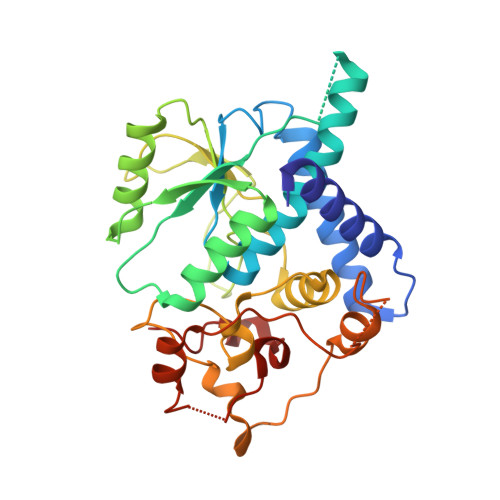
Crystal Structure of Yeast Fad Synthetase (Fad1) in Complex with Fad.

Leulliot, N.Blondeau, K.Keller, J.Ulryck, N.Quevillon-Cheruel, S.Van Tilbeurgh, H.

(2010) J Mol Biology 398: 641

  • DOI: https://doi.org/10.1016/j.jmb.2010.03.040
  • Primary Citation of Related Structures:  
    2WSI

  • PubMed Abstract: 

    Flavin adenine dinucleotide (FAD) synthetase is an essential enzyme responsible for the synthesis of FAD by adenylation of riboflavin monophosphate (FMN). We have solved the 1.9 A resolution structure of Fad1, the yeast FAD synthetase, in complex with the FAD product in the active site. The structure of Fad1 shows it to be a member of the PP-ATPase superfamily. Important conformational differences in the two motifs involved in binding the phosphate moieties of FAD compared to the Candida glabrata FMNT ortholog suggests that this loop is dynamic and undergoes substantial conformational changes during its catalytic cycle.


  • Organizational Affiliation

    Institut de Biochimie et de Biophysique Moléculaire et Cellulaire, CNRS-UMR8619, Université de Paris-Sud, Bât 430, 91405 Orsay Cedex, France.


Macromolecules
Find similar proteins by:  (by identity cutoff)  |  3D Structure
Entity ID: 1
MoleculeChains Sequence LengthOrganismDetailsImage
FAD SYNTHETASE306Saccharomyces cerevisiaeMutation(s): 0 
EC: 2.7.7.2
UniProt
Find proteins for P38913 (Saccharomyces cerevisiae (strain ATCC 204508 / S288c))
Explore P38913 
Go to UniProtKB:  P38913
Entity Groups  
Sequence Clusters30% Identity50% Identity70% Identity90% Identity95% Identity100% Identity
UniProt GroupP38913
Sequence Annotations
Expand
  • Reference Sequence
Experimental Data & Validation

Experimental Data

  • Method: X-RAY DIFFRACTION
  • Resolution: 1.90 Å
  • R-Value Free:  0.231 (Depositor), 0.220 (DCC) 
  • R-Value Work:  0.174 (Depositor), 0.170 (DCC) 
  • R-Value Observed: 0.177 (Depositor) 
Space Group: C 1 2 1
Unit Cell:
Length ( Å )Angle ( ˚ )
a = 101.871α = 90
b = 36.861β = 92.35
c = 79.231γ = 90
Software Package:
Software NamePurpose
PHENIXrefinement
PHENIXphasing

Structure Validation

View Full Validation Report


Ligand Structure Quality Assessment 

Created with Raphaël 2.3.0Worse 01 BetterLigand structure goodness of fit to experimental dataBest fitted FADClick on this verticalbar to view details

Entry History 

Deposition Data

Revision History  (Full details and data files)

  • Version 1.0: 2010-05-26
    Type: Initial release
  • Version 1.1: 2011-05-08
    Changes: Version format compliance
  • Version 1.2: 2011-07-13
    Changes: Version format compliance
  • Version 1.3: 2024-05-08
    Changes: Data collection, Database references, Derived calculations, Other