2WLT

The crystal structure of Helicobacter pylori L-asparaginase at 1.4 A resolution


Experimental Data Snapshot

  • Method: X-RAY DIFFRACTION
  • Resolution: 1.40 Å
  • R-Value Free: 0.169 
  • R-Value Observed: 0.131 

wwPDB Validation   3D Report Full Report


This is version 1.4 of the entry. See complete history


Literature

Structure of Helicobacter Pylori L-Asparaginase at 1.4 A Resolution

Dhavala, P.Papageorgiou, A.C.

(2009) Acta Crystallogr D Biol Crystallogr 65: 1253

  • DOI: https://doi.org/10.1107/S0907444909038244
  • Primary Citation of Related Structures:  
    2WLT, 2WT4

  • PubMed Abstract: 

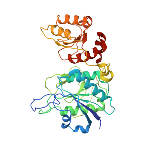
    Bacterial L-asparaginases have been used in the treatment of childhood acute lymphoblastic leukaemia for over 30 years. Their therapeutic effect is based on their ability to catalyze the conversion of L-asparagine, an essential amino acid in certain tumours, to L-aspartic acid and ammonia. Two L-asparaginases, one from Escherichia coli and the other from Erwinia chrysanthemi, have been widely employed in clinical practice as anti-leukaemia drugs. However, L-asparaginases are also able to cause severe side effects owing to their intrinsic glutaminase activity. Helicobacter pylori L-asparaginase (HpA) has been reported to have negligible glutaminase activity. To gain insight into the properties of HpA, its crystal structure in the presence of L-aspartate was determined to 1.4 A resolution, which is one of the highest resolutions obtained for an L-asparaginase structure. The final structure has an R(cryst) of 12.6% (R(free) = 16.9%) with good stereochemistry. A detailed analysis of the active site showed major differences in the active-site flexible loop and in the 286-297 loop from the second subunit, which is involved in active-site formation. Accordingly, Glu289, Asn255 and Gln63 are suggested to play roles in modulating the accessibility of the active site. Overall, the structural comparison revealed that HpA has greater structural similarity to E. coli L-asparaginase than to any other L-asparaginase, including Er. carotovora L-asparaginase, despite the fact that the latter is also characterized by low glutaminase activity.


  • Organizational Affiliation

    Turku Centre for Biotechnology, University of Turku and Abo Akademi University, Finland.


Macromolecules
Find similar proteins by:  (by identity cutoff)  |  3D Structure
Entity ID: 1
MoleculeChains Sequence LengthOrganismDetailsImage
L-ASPARAGINASE332Helicobacter pyloriMutation(s): 0 
EC: 3.5.1.1
UniProt
Find proteins for Q9ZLB9 (Helicobacter pylori (strain J99 / ATCC 700824))
Explore Q9ZLB9 
Go to UniProtKB:  Q9ZLB9
Entity Groups  
Sequence Clusters30% Identity50% Identity70% Identity90% Identity95% Identity100% Identity
UniProt GroupQ9ZLB9
Sequence Annotations
Expand
  • Reference Sequence
Small Molecules
Ligands 1 Unique
IDChains Name / Formula / InChI Key2D Diagram3D Interactions
ASP
Query on ASP

Download Ideal Coordinates CCD File 
B [auth A]ASPARTIC ACID
C4 H7 N O4
CKLJMWTZIZZHCS-REOHCLBHSA-N
Experimental Data & Validation

Experimental Data

  • Method: X-RAY DIFFRACTION
  • Resolution: 1.40 Å
  • R-Value Free: 0.169 
  • R-Value Observed: 0.131 
  • Space Group: I 2 2 2
Unit Cell:
Length ( Å )Angle ( ˚ )
a = 63.559α = 90
b = 94.796β = 90
c = 100.342γ = 90
Software Package:
Software NamePurpose
SHELXL-97refinement
HKLdata reduction
HKLdata scaling
PHASERphasing

Structure Validation

View Full Validation Report


Entry History 

Deposition Data

Revision History  (Full details and data files)

  • Version 1.0: 2009-11-24
    Type: Initial release
  • Version 1.1: 2012-05-30
    Changes: Atomic model, Database references, Derived calculations, Other, Version format compliance
  • Version 1.2: 2019-05-22
    Changes: Data collection, Other, Refinement description
  • Version 1.3: 2019-07-24
    Changes: Data collection
  • Version 1.4: 2023-12-13
    Changes: Data collection, Database references, Derived calculations, Other, Refinement description