2WL8

X-ray crystal structure of Pex19p


Experimental Data Snapshot

  • Method: X-RAY DIFFRACTION
  • Resolution: 2.05 Å
  • R-Value Free: 0.239 
  • R-Value Work: 0.199 
  • R-Value Observed: 0.201 

wwPDB Validation   3D Report Full Report


This is version 1.3 of the entry. See complete history


Literature

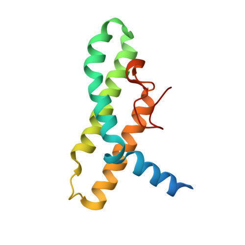
The Peroxisomal Receptor Pex19P Forms a Helical Mpts Recognition Domain.

Schueller, N.Holton, S.J.Fodor, K.Milewski, M.Konarev, P.Stanley, W.A.Wolf, J.Erdmann, R.Schliebs, W.Song, Y.H.Wilmanns, M.

(2010) EMBO J 29: 2491

  • DOI: https://doi.org/10.1038/emboj.2010.115
  • Primary Citation of Related Structures:  
    2WL8

  • PubMed Abstract: 

    The protein Pex19p functions as a receptor and chaperone of peroxisomal membrane proteins (PMPs). The crystal structure of the folded C-terminal part of the receptor reveals a globular domain that displays a bundle of three long helices in an antiparallel arrangement. Complementary functional experiments, using a range of truncated Pex19p constructs, show that the structured alpha-helical domain binds PMP-targeting signal (mPTS) sequences with about 10 muM affinity. Removal of a conserved N-terminal helical segment from the mPTS recognition domain impairs the ability for mPTS binding, indicating that it forms part of the mPTS-binding site. Pex19p variants with mutations in the same sequence segment abolish correct cargo import. Our data indicate a divided N-terminal and C-terminal structural arrangement in Pex19p, which is reminiscent of a similar division in the Pex5p receptor, to allow separation of cargo-targeting signal recognition and additional functions.


  • Organizational Affiliation

    EMBL c/o DESY, Notkestrasse 85, Hamburg, Germany.


Macromolecules
Find similar proteins by:  (by identity cutoff)  |  3D Structure
Entity ID: 1
MoleculeChains Sequence LengthOrganismDetailsImage
PEROXISOMAL BIOGENESIS FACTOR 19
A, B, C, D
126Homo sapiensMutation(s): 0 
Membrane Entity: Yes 
UniProt & NIH Common Fund Data Resources
Find proteins for P40855 (Homo sapiens)
Explore P40855 
Go to UniProtKB:  P40855
PHAROS:  P40855
GTEx:  ENSG00000162735 
Entity Groups  
Sequence Clusters30% Identity50% Identity70% Identity90% Identity95% Identity100% Identity
UniProt GroupP40855
Sequence Annotations
Expand
  • Reference Sequence
Experimental Data & Validation

Experimental Data

  • Method: X-RAY DIFFRACTION
  • Resolution: 2.05 Å
  • R-Value Free: 0.239 
  • R-Value Work: 0.199 
  • R-Value Observed: 0.201 
  • Space Group: P 21 21 21
Unit Cell:
Length ( Å )Angle ( ˚ )
a = 67.15α = 90
b = 91.12β = 90
c = 122.287γ = 90
Software Package:
Software NamePurpose
REFMACrefinement
MOSFLMdata reduction
TRUNCATEdata scaling

Structure Validation

View Full Validation Report


Entry History 

Deposition Data

Revision History  (Full details and data files)

  • Version 1.0: 2010-06-23
    Type: Initial release
  • Version 1.1: 2011-07-13
    Changes: Advisory, Version format compliance
  • Version 1.2: 2011-09-21
    Changes: Database references
  • Version 1.3: 2019-07-24
    Changes: Data collection