2WDR

E. coli succinate:quinone oxidoreductase (SQR) with pentachlorophenol bound


Experimental Data Snapshot

  • Method: X-RAY DIFFRACTION
  • Resolution: 3.20 Å
  • R-Value Free: 0.228 
  • R-Value Work: 0.197 
  • R-Value Observed: 0.199 

wwPDB Validation   3D Report Full Report


Ligand Structure Quality Assessment 


This is version 1.2 of the entry. See complete history


Literature

Structure of Escherichia Coli Succinate:Quinone Oxidoreductase with an Occupied and Empty Quinone- Binding Site.

Ruprecht, J.Yankovskaya, V.Maklashina, E.Iwata, S.Cecchini, G.

(2009) J Biol Chem 284: 29836

  • DOI: https://doi.org/10.1074/jbc.M109.010058
  • Primary Citation of Related Structures:  
    2WDQ, 2WDR, 2WDV

  • PubMed Abstract: 

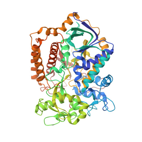
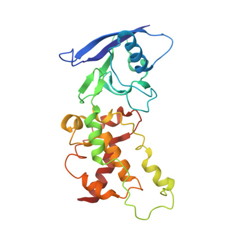
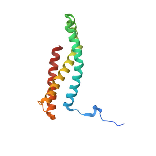
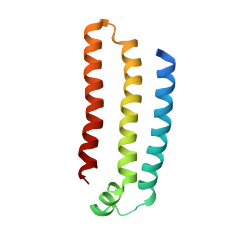
    Three new structures of Escherichia coli succinate-quinone oxidoreductase (SQR) have been solved. One with the specific quinone-binding site (Q-site) inhibitor carboxin present has been solved at 2.4 A resolution and reveals how carboxin inhibits the Q-site. The other new structures are with the Q-site inhibitor pentachlorophenol and with an empty Q-site. These structures reveal important details unresolved in earlier structures. Comparison of the new SQR structures shows how subtle rearrangements of the quinone-binding site accommodate the different inhibitors. The position of conserved water molecules near the quinone binding pocket leads to a reassessment of possible water-mediated proton uptake networks that complete reduction of ubiquinone. The dicarboxylate-binding site in the soluble domain of SQR is highly similar to that seen in high resolution structures of avian SQR (PDB 2H88) and soluble flavocytochrome c (PDB 1QJD) showing mechanistically significant structural features conserved across prokaryotic and eukaryotic SQRs.


  • Organizational Affiliation

    Membrane Protein Crystallography Group, Molecular Biosciences Division, Imperial College, London SW72AZ, United Kingdom.


Macromolecules
Find similar proteins by:  (by identity cutoff)  |  3D Structure
Entity ID: 1
MoleculeChains Sequence LengthOrganismDetailsImage
SUCCINATE DEHYDROGENASE FLAVOPROTEIN SUBUNIT
A, E, I
588Escherichia coliMutation(s): 0 
EC: 1.3.5.1 (PDB Primary Data), 1.3.99.1 (PDB Primary Data)
Membrane Entity: Yes 
UniProt
Find proteins for P0AC41 (Escherichia coli (strain K12))
Explore P0AC41 
Go to UniProtKB:  P0AC41
Entity Groups  
Sequence Clusters30% Identity50% Identity70% Identity90% Identity95% Identity100% Identity
UniProt GroupP0AC41
Sequence Annotations
Expand
  • Reference Sequence
Find similar proteins by:  (by identity cutoff)  |  3D Structure
Entity ID: 2
MoleculeChains Sequence LengthOrganismDetailsImage
SUCCINATE DEHYDROGENASE IRON-SULFUR SUBUNIT
B, F, J
238Escherichia coliMutation(s): 0 
EC: 1.3.5.1 (PDB Primary Data), 1.3.99.1 (PDB Primary Data)
Membrane Entity: Yes 
UniProt
Find proteins for P07014 (Escherichia coli (strain K12))
Explore P07014 
Go to UniProtKB:  P07014
Entity Groups  
Sequence Clusters30% Identity50% Identity70% Identity90% Identity95% Identity100% Identity
UniProt GroupP07014
Sequence Annotations
Expand
  • Reference Sequence
Find similar proteins by:  (by identity cutoff)  |  3D Structure
Entity ID: 3
MoleculeChains Sequence LengthOrganismDetailsImage
SUCCINATE DEHYDROGENASE CYTOCHROME B556 SUBUNIT
C, G, K
129Escherichia coliMutation(s): 0 
EC: 1.3.5.1
Membrane Entity: Yes 
UniProt
Find proteins for P69054 (Escherichia coli (strain K12))
Explore P69054 
Go to UniProtKB:  P69054
Entity Groups  
Sequence Clusters30% Identity50% Identity70% Identity90% Identity95% Identity100% Identity
UniProt GroupP69054
Sequence Annotations
Expand
  • Reference Sequence
Find similar proteins by:  (by identity cutoff)  |  3D Structure
Entity ID: 4
MoleculeChains Sequence LengthOrganismDetailsImage
SUCCINATE DEHYDROGENASE HYDROPHOBIC MEMBRANE ANCHOR SUBUNIT
D, H, L
115Escherichia coliMutation(s): 0 
EC: 1.3.5.1
Membrane Entity: Yes 
UniProt
Find proteins for P0AC44 (Escherichia coli (strain K12))
Explore P0AC44 
Go to UniProtKB:  P0AC44
Entity Groups  
Sequence Clusters30% Identity50% Identity70% Identity90% Identity95% Identity100% Identity
UniProt GroupP0AC44
Sequence Annotations
Expand
  • Reference Sequence
Small Molecules
Ligands 8 Unique
IDChains Name / Formula / InChI Key2D Diagram3D Interactions
FAD
Query on FAD

Download Ideal Coordinates CCD File 
CA [auth I],
M [auth A],
U [auth E]
FLAVIN-ADENINE DINUCLEOTIDE
C27 H33 N9 O15 P2
VWWQXMAJTJZDQX-UYBVJOGSSA-N
HEM
Query on HEM

Download Ideal Coordinates CCD File 
AA [auth G],
IA [auth K],
S [auth C]
PROTOPORPHYRIN IX CONTAINING FE
C34 H32 Fe N4 O4
KABFMIBPWCXCRK-RGGAHWMASA-L
SF4
Query on SF4

Download Ideal Coordinates CCD File 
GA [auth J],
Q [auth B],
Y [auth F]
IRON/SULFUR CLUSTER
Fe4 S4
LJBDFODJNLIPKO-UHFFFAOYSA-N
F3S
Query on F3S

Download Ideal Coordinates CCD File 
HA [auth J],
R [auth B],
Z [auth F]
FE3-S4 CLUSTER
Fe3 S4
FCXHZBQOKRZXKS-UHFFFAOYSA-N
PCI
Query on PCI

Download Ideal Coordinates CCD File 
BA [auth G],
JA [auth K],
T [auth C]
PENTACHLOROPHENOL
C6 H Cl5 O
IZUPBVBPLAPZRR-UHFFFAOYSA-N
FES
Query on FES

Download Ideal Coordinates CCD File 
FA [auth J],
P [auth B],
X [auth F]
FE2/S2 (INORGANIC) CLUSTER
Fe2 S2
NIXDOXVAJZFRNF-UHFFFAOYSA-N
TEO
Query on TEO

Download Ideal Coordinates CCD File 
DA [auth I],
N [auth A],
V [auth E]
MALATE LIKE INTERMEDIATE
C4 H4 O5
QFBHYOKSQPPXHZ-UWTATZPHSA-L
NA
Query on NA

Download Ideal Coordinates CCD File 
EA [auth I],
O [auth A],
W [auth E]
SODIUM ION
Na
FKNQFGJONOIPTF-UHFFFAOYSA-N
Experimental Data & Validation

Experimental Data

  • Method: X-RAY DIFFRACTION
  • Resolution: 3.20 Å
  • R-Value Free: 0.228 
  • R-Value Work: 0.197 
  • R-Value Observed: 0.199 
  • Space Group: P 21 21 21
Unit Cell:
Length ( Å )Angle ( ˚ )
a = 119.955α = 90
b = 186.128β = 90
c = 204.034γ = 90
Software Package:
Software NamePurpose
REFMACrefinement
XDSdata reduction
SCALAdata scaling
REFMACphasing

Structure Validation

View Full Validation Report


Ligand Structure Quality Assessment 


Entry History 

Deposition Data

Revision History  (Full details and data files)

  • Version 1.0: 2009-08-25
    Type: Initial release
  • Version 1.1: 2011-07-13
    Changes: Advisory, Version format compliance
  • Version 1.2: 2015-02-04
    Changes: Atomic model, Derived calculations, Non-polymer description, Other