2WC1 | pdb_00002wc1

Three-dimensional Structure of the Nitrogen Fixation Flavodoxin (NifF) from Rhodobacter capsulatus at 2.2 A


Experimental Data Snapshot

  • Method: X-RAY DIFFRACTION
  • Resolution: 2.17 Å
  • R-Value Free: 
    0.267 (Depositor), 0.251 (DCC) 
  • R-Value Work: 
    0.246 (Depositor), 0.247 (DCC) 
  • R-Value Observed: 
    0.246 (Depositor) 

Starting Model: experimental
View more details

wwPDB Validation 3D Report Full Report

Validation slider image for 2WC1

Ligand Structure Quality Assessment 


This is version 1.2 of the entry. See complete history

Literature

Structural and Phylogenetic Analysis of Rhodobacter Capsulatus Niff: Uncovering General Features of Nitrogen-Fixation (Nif)-Flavodoxins.

Perez-Dorado, I.Bortolotti, A.Cortez, N.Hermoso, J.A.

(2013) Int J Mol Sci 14: 1152

  • DOI: https://doi.org/10.3390/ijms14011152
  • Primary Citation Related Structures: 
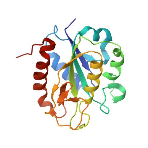
    2WC1

  • PubMed Abstract: 

    Analysis of the crystal structure of NifF from Rhodobacter capsulatus and its homologues reported so far reflects the existence of unique structural features in nif flavodoxins: a leucine at the re face of the isoalloxazine, an eight-residue insertion at the C-terminus of the 50's loop and a remarkable difference in the electrostatic potential surface with respect to non-nif flavodoxins. A phylogenetic study on 64 sequences from 52 bacterial species revealed four clusters, including different functional prototypes, correlating the previously defined as "short-chain" with the firmicutes flavodoxins and the "long-chain" with gram-negative species. The comparison of Rhodobacter NifF structure with other bacterial flavodoxin prototypes discloses the concurrence of specific features of these functional electron donors to nitrogenase.


  • Organizational Affiliation
    • Department of Crystallography and Structural Biology, Institute of Physical-Chemistry "Rocasolano", CSIC, Serrano 119, Madrid 28006, Spain. xjuan@iqfr.csic.es.

Macromolecule Content 

  • Total Structure Weight: 20.29 kDa 
  • Atom Count: 1,507 
  • Modeled Residue Count: 182 
  • Deposited Residue Count: 182 
  • Unique protein chains: 1

Macromolecules

Find similar proteins by:|  3D Structure
Entity ID: 1
MoleculeChains  Sequence LengthOrganismDetailsImage
FLAVODOXIN182Rhodobacter capsulatusMutation(s): 0 
UniProt
Find proteins for P52967 (Rhodobacter capsulatus (strain ATCC BAA-309 / NBRC 16581 / SB1003))
Explore P52967 
Go to UniProtKB:  P52967
Entity Groups
Sequence Clusters30% Identity50% Identity70% Identity90% Identity95% Identity100% Identity
UniProt GroupP52967
Sequence Annotations
Expand
Reference Sequence

Small Molecules

Ligands 1 Unique
IDChains Name / Formula / InChI Key2D Diagram3D Interactions
FMN

Query on FMN


Download:Ideal Coordinates CCD File
B [auth A]FLAVIN MONONUCLEOTIDE
C17 H21 N4 O9 P
FVTCRASFADXXNN-SCRDCRAPSA-N

Experimental Data & Validation

Experimental Data

  • Method: X-RAY DIFFRACTION
  • Resolution: 2.17 Å
  • R-Value Free:  0.267 (Depositor), 0.251 (DCC) 
  • R-Value Work:  0.246 (Depositor), 0.247 (DCC) 
  • R-Value Observed: 0.246 (Depositor) 
Space Group: P 41 21 2
Unit Cell:
Length ( Å )Angle ( ˚ )
a = 66.4α = 90
b = 66.4β = 90
c = 121.32γ = 90
Software Package:
Software NamePurpose
CNSrefinement
MOSFLMdata reduction
SCALAdata scaling
MOLREPphasing

Structure Validation

View Full Validation Report


Ligand Structure Quality Assessment 


Entry History 

Deposition Data

Revision History  (Full details and data files)

  • Version 1.0: 2010-04-21
    Type: Initial release
  • Version 1.1: 2014-07-30
    Changes: Database references
  • Version 1.2: 2023-12-13
    Changes: Data collection, Database references, Derived calculations, Other, Refinement description