2WB2

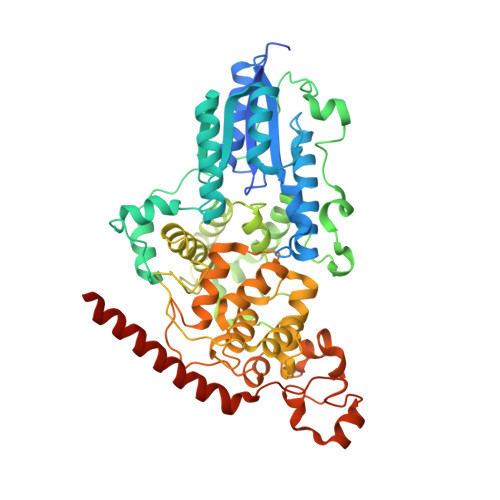
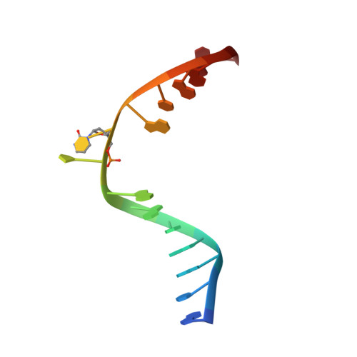
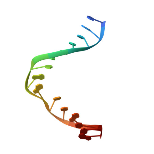
Drosophila Melanogaster (6-4) Photolyase Bound To double stranded Dna containing a T(6-4)C Photolesion


Experimental Data Snapshot

  • Method: X-RAY DIFFRACTION
  • Resolution: 2.95 Å
  • R-Value Free: 0.243 
  • R-Value Work: 0.204 
  • R-Value Observed: 0.206 

wwPDB Validation   3D Report Full Report


Ligand Structure Quality Assessment 


This is version 2.0 of the entry. See complete history


Literature

Crystal Structure of the T(6-4)C Lesion in Complex with a (6-4) DNA Photolyase and Repair of Uv- Induced (6-4) and Dewar Photolesions.

Glas, A.F.Schneider, S.Maul, M.J.Hennecke, U.Carell, T.

(2009) Chemistry 15: 10387

  • DOI: https://doi.org/10.1002/chem.200901004
  • Primary Citation of Related Structures:  
    2WB2

  • PubMed Abstract: 

    UV-light irradiation induces the formation of highly mutagenic lesions in DNA, such as cis-syn cyclobutane pyrimidine dimers (CPD photoproducts), pyrimidine(6-4)pyrimidone photoproducts ((6-4) photoproducts) and their Dewar valence isomers ((Dew) photoproducts). Here we describe the synthesis of defined DNA strands containing these lesions by direct irradiation. We show that all lesions are efficiently repaired except for the T(Dew)T lesion, which cannot be cleaved by the repair enzyme under our conditions. A crystal structure of a T(6-4)C lesion containing DNA duplex in complex with the (6-4) photolyase from Drosophila melanogaster provides insight into the molecular recognition event of a cytosine derived photolesion for the first time. In light of the previously postulated repair mechanism, which involves rearrangement of the (6-4) lesions into strained four-membered ring repair intermediates, it is surprising that the not rearranged T(6-4)C lesion is observed in the active site. The structure, therefore, provides additional support for the newly postulated repair mechanism that avoids this rearrangement step and argues for a direct electron injection into the lesion as the first step of the repair reaction performed by (6-4) DNA photolyases.


  • Organizational Affiliation

    Department for Chemistry and Biochemistry, Ludwig-Maximilians University, Butenandtstr. 5-13, 81377 Munich, Germany.


Macromolecules

Find similar proteins by:  (by identity cutoff)  |  3D Structure
Entity ID: 1
MoleculeChains Sequence LengthOrganismDetailsImage
PHOTOLYASE543Drosophila melanogasterMutation(s): 0 
EC: 4.1.99.3
UniProt
Find proteins for Q8SXK5 (Drosophila melanogaster)
Explore Q8SXK5 
Go to UniProtKB:  Q8SXK5
Entity Groups  
Sequence Clusters30% Identity50% Identity70% Identity90% Identity95% Identity100% Identity
UniProt GroupQ8SXK5
Sequence Annotations
Expand
  • Reference Sequence

Find similar nucleic acids by:  Sequence   |   3D Structure  

Entity ID: 2
MoleculeChains LengthOrganismImage
5'-D(*AP*CP*AP*GP*CP*GP*GP*64PP*ZP*GP*CP*AP *GP*GP*T)-3'B [auth C]15synthetic construct
Sequence Annotations
Expand
  • Reference Sequence

Find similar nucleic acids by:  Sequence   |   3D Structure  

Entity ID: 3
MoleculeChains LengthOrganismImage
5'-D(*TP*AP*CP*CP*TP*GP*CP*GP*AP*CP*CP*GP*CP*TP*G)-3'C [auth D]15synthetic construct
Sequence Annotations
Expand
  • Reference Sequence
Small Molecules
Ligands 1 Unique
IDChains Name / Formula / InChI Key2D Diagram3D Interactions
FAD
Query on FAD

Download Ideal Coordinates CCD File 
D [auth A]FLAVIN-ADENINE DINUCLEOTIDE
C27 H33 N9 O15 P2
VWWQXMAJTJZDQX-UYBVJOGSSA-N
Experimental Data & Validation

Experimental Data

  • Method: X-RAY DIFFRACTION
  • Resolution: 2.95 Å
  • R-Value Free: 0.243 
  • R-Value Work: 0.204 
  • R-Value Observed: 0.206 
  • Space Group: P 21 21 21
Unit Cell:
Length ( Å )Angle ( ˚ )
a = 87.55α = 90
b = 89β = 90
c = 91.67γ = 90
Software Package:
Software NamePurpose
REFMACrefinement
XDSdata reduction
SCALAdata scaling
PHASERphasing

Structure Validation

View Full Validation Report


Ligand Structure Quality Assessment 


Entry History 

Deposition Data

Revision History  (Full details and data files)

  • Version 1.0: 2009-03-17
    Type: Initial release
  • Version 1.1: 2011-07-13
    Changes: Advisory, Version format compliance
  • Version 1.2: 2011-10-26
    Changes: Database references, Source and taxonomy, Structure summary
  • Version 1.3: 2019-05-29
    Changes: Data collection, Derived calculations, Experimental preparation, Other, Source and taxonomy
  • Version 1.4: 2019-08-07
    Changes: Data collection, Experimental preparation
  • Version 1.5: 2019-10-16
    Changes: Data collection, Other
  • Version 2.0: 2023-12-13
    Changes: Data collection, Database references, Derived calculations, Polymer sequence, Refinement description