


Crystal Structure of Human Zinc-Binding Alcohol Dehydrogenase 1
Shafqat, N., Yue, W.W., Niesen, F., Oppermann, U.To be published.
Experimental Data Snapshot
Starting Model: experimental
View more details
Entity ID: 1 | |||||
|---|---|---|---|---|---|
| Molecule | Chains | Sequence Length | Organism | Details | Image |
| PROSTAGLANDIN REDUCTASE 2 | 357 | Homo sapiens | Mutation(s): 0 EC: 1.3.1.48 | 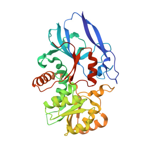 | |
UniProt & NIH Common Fund Data Resources | |||||
PHAROS: Q8N8N7 GTEx: ENSG00000140043 | |||||
Entity Groups | |||||
| Sequence Clusters | 30% Identity50% Identity70% Identity90% Identity95% Identity100% Identity | ||||
| UniProt Group | Q8N8N7 | ||||
Sequence AnnotationsExpand | |||||
Reference Sequence | |||||
| Ligands 6 Unique | |||||
|---|---|---|---|---|---|
| ID | Chains | Name / Formula / InChI Key | 2D Diagram | 3D Interactions | |
| NAP Download:Ideal Coordinates CCD File | C [auth A], J [auth B] | NADP NICOTINAMIDE-ADENINE-DINUCLEOTIDE PHOSPHATE C21 H28 N7 O17 P3 XJLXINKUBYWONI-NNYOXOHSSA-N |  | ||
| P1Z Download:Ideal Coordinates CCD File | D [auth A] E [auth A] F [auth A] K [auth B] L [auth B] | 4-BUTYL-1,2-DIPHENYL-PYRAZOLIDINE-3,5-DIONE C19 H20 N2 O2 VYMDGNCVAMGZFE-UHFFFAOYSA-N |  | ||
| LMR Download:Ideal Coordinates CCD File | Q [auth B] | (2S)-2-hydroxybutanedioic acid C4 H6 O5 BJEPYKJPYRNKOW-REOHCLBHSA-N |  | ||
| PO4 Download:Ideal Coordinates CCD File | G [auth A], H [auth A], I [auth A], O [auth B], P [auth B] | PHOSPHATE ION O4 P NBIIXXVUZAFLBC-UHFFFAOYSA-K |  | ||
| CL Download:Ideal Coordinates CCD File | R [auth B] | CHLORIDE ION Cl VEXZGXHMUGYJMC-UHFFFAOYSA-M |  | ||
| NA Download:Ideal Coordinates CCD File | S [auth B] | SODIUM ION Na FKNQFGJONOIPTF-UHFFFAOYSA-N |  | ||
| Length ( Å ) | Angle ( ˚ ) |
|---|---|
| a = 141.014 | α = 90 |
| b = 82.793 | β = 99.77 |
| c = 69.097 | γ = 90 |
| Software Name | Purpose |
|---|---|
| REFMAC | refinement |
| MOSFLM | data reduction |
| SCALA | data scaling |
| PHASER | phasing |


