2W59 | pdb_00002w59

STRUCTURE OF AN AVIAN IGY-FC 3-4 FRAGMENT


Experimental Data Snapshot

  • Method: X-RAY DIFFRACTION
  • Resolution: 1.75 Å
  • R-Value Free: 
    0.196 (Depositor), 0.297 (DCC) 
  • R-Value Work: 
    0.169 (Depositor) 
  • R-Value Observed: 
    0.171 (Depositor) 

Starting Model: experimental
View more details

wwPDB Validation 3D Report Full Report

Validation slider image for 2W59

This is version 2.2 of the entry. See complete history

Literature

The Crystal Structure of an Avian Igy-Fc Fragment Reveals Conservation with Both Mammalian Igg and Ige.

Taylor, A.I.Fabiane, S.M.Sutton, B.J.Calvert, R.A.

(2009) Biochemistry 48: 558

  • DOI: https://doi.org/10.1021/bi8019993
  • Primary Citation Related Structures: 
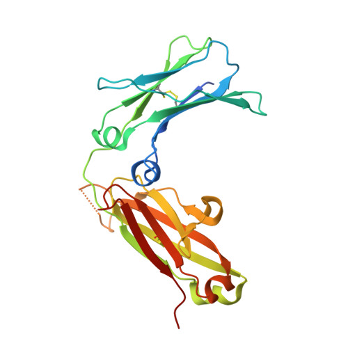
    2W59

  • PubMed Abstract: 

    Avian IgY is closely related to an ancestor of both mammalian IgG and IgE and thus provides insights into the evolution of antibody structure and function. A recombinant fragment of IgY-Fc consisting of a dimer of the Cupsilon3 and Cupsilon4 domains, Fcupsilon3-4, was expressed and crystallized and its X-ray structure determined to 1.75 A resolution. Fcupsilon3-4 is the only nonmammalian Fc fragment structure determined to date and provides the first structural evidence for an ancient origin of antibody architecture. The Fcupsilon3-4 structure reveals features common to both IgE-Fc and IgG-Fc, and the implications for IgY binding to its receptor are discussed.


  • Organizational Affiliation
    • Randall Division of Cell and Molecular Biophysics, King's College London, and MRC-Asthma UK Centre in Allergic Mechanisms of Asthma, New Hunt's House, Guy's Campus, London SE1 1UL, United Kingdom.

Macromolecule Content 

  • Total Structure Weight: 52.71 kDa 
  • Atom Count: 4,072 
  • Modeled Residue Count: 429 
  • Deposited Residue Count: 462 
  • Unique protein chains: 1

Macromolecules

Find similar proteins by:|  3D Structure
Entity ID: 1
MoleculeChains  Sequence LengthOrganismDetailsImage
IGY FCU3-4
A, B
231Gallus gallusMutation(s): 0 

Oligosaccharides

Help  
Entity ID: 2
MoleculeChains Length2D Diagram GlycosylationD Interactions
alpha-D-mannopyranose-(1-4)-2-acetamido-2-deoxy-beta-D-glucopyranose-(1-4)-2-acetamido-2-deoxy-beta-D-glucopyranose
C, D
3N-Glycosylation
Glycosylation Resources
GlyTouCan: G62182OO
GlyCosmos: G62182OO
GlyGen: G62182OO

Experimental Data & Validation

Experimental Data

  • Method: X-RAY DIFFRACTION
  • Resolution: 1.75 Å
  • R-Value Free:  0.196 (Depositor), 0.297 (DCC) 
  • R-Value Work:  0.169 (Depositor) 
  • R-Value Observed: 0.171 (Depositor) 
Space Group: P 21 21 21
Unit Cell:
Length ( Å )Angle ( ˚ )
a = 68.481α = 90
b = 80.14β = 90
c = 99.135γ = 90
Software Package:
Software NamePurpose
PHENIXrefinement
MOSFLMdata reduction
SCALAdata scaling
PHASERphasing

Structure Validation

View Full Validation Report


Entry History 

Deposition Data

Revision History  (Full details and data files)

  • Version 1.0: 2009-01-13
    Type: Initial release
  • Version 1.1: 2011-05-08
    Changes: Version format compliance
  • Version 1.2: 2011-07-13
    Changes: Version format compliance
  • Version 1.3: 2017-07-12
    Changes: Advisory
  • Version 2.0: 2020-07-29
    Type: Remediation
    Reason: Carbohydrate remediation
    Changes: Advisory, Atomic model, Data collection, Derived calculations, Other, Structure summary
  • Version 2.1: 2023-12-13
    Changes: Data collection, Database references, Refinement description, Structure summary
  • Version 2.2: 2024-10-16
    Changes: Structure summary