2W42 | pdb_00002w42

THE STRUCTURE OF A PIWI PROTEIN FROM ARCHAEOGLOBUS FULGIDUS COMPLEXED WITH A 16NT DNA DUPLEX.


Experimental Data Snapshot

  • Method: X-RAY DIFFRACTION
  • Resolution: 1.90 Å
  • R-Value Free: 
    0.243 (Depositor), 0.241 (DCC) 
  • R-Value Work: 
    0.205 (Depositor), 0.204 (DCC) 
  • R-Value Observed: 
    0.207 (Depositor) 

Starting Model: experimental
View more details

wwPDB Validation 3D Report Full Report

Validation slider image for 2W42

This is version 1.3 of the entry. See complete history

Literature

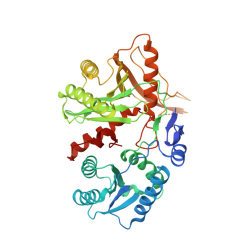
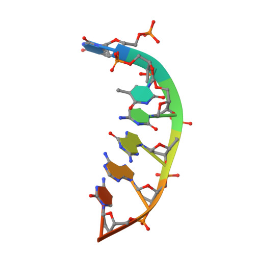
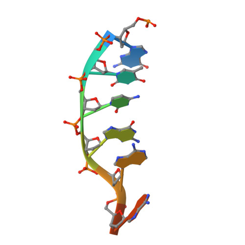
Enhancement of the Seed-Target Recognition Step in RNA Silencing by a Piwi-Mid Domain Protein

Parker, J.S.Parizotto, E.A.Wang, M.Roe, S.M.Barford, D.

(2009) Mol Cell 33: 204

  • DOI: https://doi.org/10.1016/j.molcel.2008.12.012
  • Primary Citation Related Structures: 
    2W42

  • PubMed Abstract: 

    Target recognition in RNA silencing is governed by the "seed sequence" of a guide RNA strand associated with the PIWI/MID domain of an Argonaute protein in RISC. Using a reconstituted in vitro target recognition system, we show that a model PIWI/MID domain protein confers position-dependent tightening and loosening of guide-strand-target interactions. Over the seed sequence, the interaction affinity is enhanced up to approximately 300-fold. Enhancement is achieved through a reduced entropy penalty for the interaction. In contrast, interactions 3' of the seed are inhibited. We quantified mismatched target recognition inside and outside the seed, revealing amplified discrimination at the third position in the seed mediated by the PIWI/MID domain. Thus, association of the guide strand with the PIWI/MID domain generates an enhanced affinity anchor site over the seed that can promote fidelity in target recognition and stabilize and guide the assembly of the active silencing complex.


  • Organizational Affiliation
    • Institute of Cancer Research, Chester Beatty Laboratories, London, UK. james.parker@bioch.ox.ac.uk

Macromolecule Content 

  • Total Structure Weight: 108.4 kDa 
  • Atom Count: 7,235 
  • Modeled Residue Count: 806 
  • Deposited Residue Count: 886 
  • Unique protein chains: 1
  • Unique nucleic acid chains: 2

Macromolecules


Find similar proteins by:|  3D Structure
Entity ID: 1
MoleculeChains  Sequence LengthOrganismDetailsImage
PUTATIVE UNCHARACTERIZED PROTEIN
A, B
427Archaeoglobus fulgidusMutation(s): 0 
UniProt
Find proteins for O28951 (Archaeoglobus fulgidus (strain ATCC 49558 / DSM 4304 / JCM 9628 / NBRC 100126 / VC-16))
Explore O28951 
Go to UniProtKB:  O28951
Entity Groups
Sequence Clusters30% Identity50% Identity70% Identity90% Identity95% Identity100% Identity
UniProt GroupO28951
Sequence Annotations
Expand
Reference Sequence
Find similar nucleic acids by:  Sequence
Entity ID: 2
MoleculeChains LengthOrganismImage
5'-D(*TP*TP*CP*GP*AP*CP*GP*CP)-3'C [auth P],
E [auth R]
8Homo sapiens
Sequence Annotations
Expand
Reference Sequence
Find similar nucleic acids by:  Sequence
Entity ID: 3
MoleculeChains LengthOrganismImage
5'-D(*GP*TP*CP*GP*AP*AP*TP*TP)-3'D [auth Q],
F [auth S]
8Homo sapiens
Sequence Annotations
Expand
Reference Sequence

Experimental Data & Validation

Experimental Data

  • Method: X-RAY DIFFRACTION
  • Resolution: 1.90 Å
  • R-Value Free:  0.243 (Depositor), 0.241 (DCC) 
  • R-Value Work:  0.205 (Depositor), 0.204 (DCC) 
  • R-Value Observed: 0.207 (Depositor) 
Space Group: P 1
Unit Cell:
Length ( Å )Angle ( ˚ )
a = 51.838α = 75.8
b = 61.384β = 75.86
c = 103.54γ = 79.47
Software Package:
Software NamePurpose
REFMACrefinement
MOSFLMdata reduction
SCALAdata scaling
PHASERphasing

Structure Validation

View Full Validation Report


Entry History 

Deposition Data

Revision History  (Full details and data files)

  • Version 1.0: 2008-12-09
    Type: Initial release
  • Version 1.1: 2011-05-08
    Changes: Version format compliance
  • Version 1.2: 2011-07-13
    Changes: Version format compliance
  • Version 1.3: 2023-12-13
    Changes: Data collection, Database references, Derived calculations, Other, Refinement description